Copyright
©The Author(s) 2016.
World J Clin Cases. Jul 16, 2016; 4(7): 177-180
Published online Jul 16, 2016. doi: 10.12998/wjcc.v4.i7.177
Published online Jul 16, 2016. doi: 10.12998/wjcc.v4.i7.177
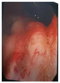
Figure 1 Endoscopic appearance of colitis cystic profunda - a polypoid lesion in the rectum covered by smooth mucosal.
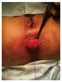
Figure 2 Intraoperative findings - excision through a vertical mucosal incision dissecting the lesion wholemeal from the submucosal layer.
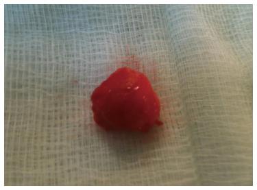
Figure 3 Operative specimen - the excised lesion was cystic containing mucous.
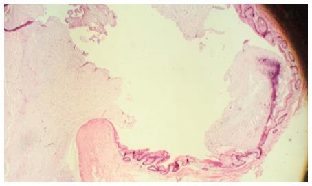
Figure 4 Hematoxylin and eosin stain of histopathology slide - this is a cyst containing inspissated mucin and lined by large bowel type benign mucinous epithelium with surrounding fibrosis.
- Citation: Ayantunde AA, Strauss C, Sivakkolunthu M, Malhotra A. Colitis cystica profunda of the rectum: An unexpected operative finding. World J Clin Cases 2016; 4(7): 177-180
- URL: https://www.wjgnet.com/2307-8960/full/v4/i7/177.htm
- DOI: https://dx.doi.org/10.12998/wjcc.v4.i7.177


