Copyright
©The Author(s) 2015.
World J Clin Cases. Apr 16, 2015; 3(4): 381-384
Published online Apr 16, 2015. doi: 10.12998/wjcc.v3.i4.381
Published online Apr 16, 2015. doi: 10.12998/wjcc.v3.i4.381
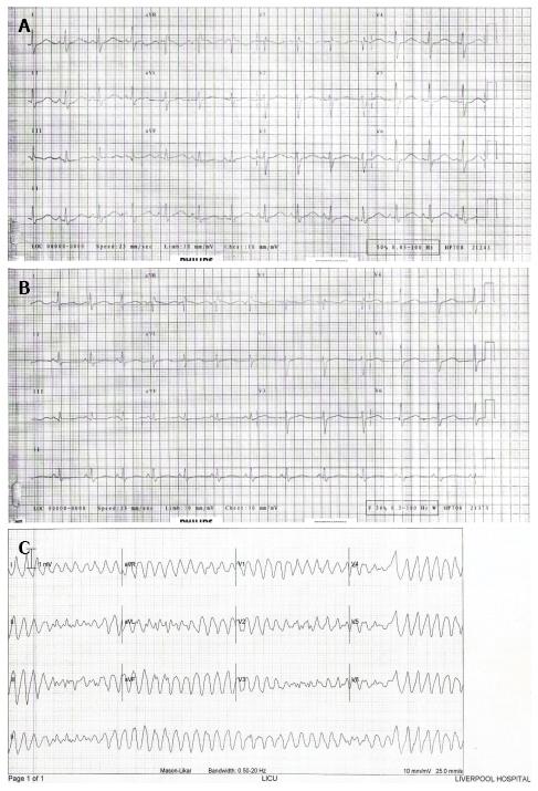
Figure 1 Electrocardiogram of AB.
A: Electrocardiogram (ECG) of AB on first presentation showed sinus rhythm, incomplete right bundle branch block (RBBB) and prolonged QTc interval (QTc 526 ms); B: ECG of AB on second presentation showed sinus rhythm, incomplete RBBB and prolonged QTc interval (QTc 505 ms); C: ECG of AB during a syncopal episode on her second admission showed torsades de pointes.
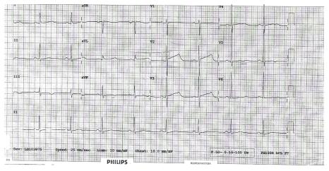
Figure 2 Electrocardiogram of CD showed sinus rhythm and prolonged QTc (QTc 566 ms).
- Citation: Choong H, Hanna I, Beran R. Importance of cardiological evaluation for first seizures. World J Clin Cases 2015; 3(4): 381-384
- URL: https://www.wjgnet.com/2307-8960/full/v3/i4/381.htm
- DOI: https://dx.doi.org/10.12998/wjcc.v3.i4.381


