Copyright
©2014 Baishideng Publishing Group Inc.
World J Clin Cases. Aug 16, 2014; 2(8): 398-401
Published online Aug 16, 2014. doi: 10.12998/wjcc.v2.i8.398
Published online Aug 16, 2014. doi: 10.12998/wjcc.v2.i8.398
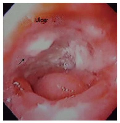
Figure 1 Endoscopic view of gastric conduit ulcer.
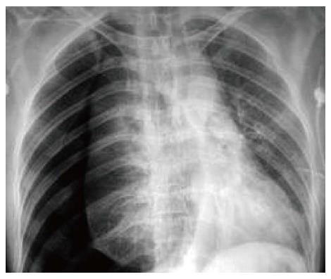
Figure 2 Chest X-ray showing right sided tension pneumothorax with mediastinal shift.
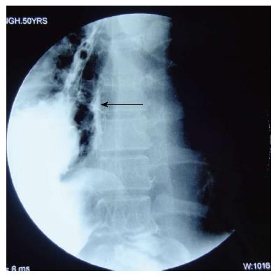
Figure 3 Oral Gastrografin study showing leak of contrast from the medial aspect of upper part of the conduit (arrow).
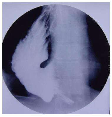
Figure 4 Repeat study after 4 wk shows no evidence of contrast leak.
- Citation: Patil N, Kaushal A, Jain A, Saluja SS, Mishra PK. Gastric conduit perforation. World J Clin Cases 2014; 2(8): 398-401
- URL: https://www.wjgnet.com/2307-8960/full/v2/i8/398.htm
- DOI: https://dx.doi.org/10.12998/wjcc.v2.i8.398


