Copyright
©2014 Baishideng Publishing Group Inc.
World J Clin Cases. Jun 16, 2014; 2(6): 219-223
Published online Jun 16, 2014. doi: 10.12998/wjcc.v2.i6.219
Published online Jun 16, 2014. doi: 10.12998/wjcc.v2.i6.219
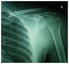
Figure 1 Anteroposterior radiograph of left shoulder showing a crescent-shaped fragment near the inferior part of the glenoid rim.
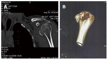
Figure 2 Further examination with computed tomography scan (A) and 3D computed tomography reconstruction (B) confirmed the diagnosis and revealed the comminution of the lesser tuberosity avulsion fracture.
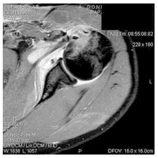
Figure 3 Magnetic resonance imaging exam of the left shoulder excluded intra-articular extension of the fracture or other tendon ruptures.
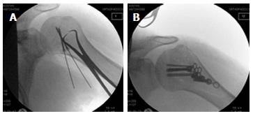
Figure 4 Intra-operative fluoroscopic images.
Initially, the bigger fragments of the lesser tuberosity were reduced using reduction forceps and fixed using cannulated headless Hebert screws (A). The smaller fragments were then reduced and stabilized under a low profile, bendable neutralization buttress plate (B).
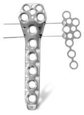
Figure 5 The 7-holes T-minus plate (right) that was used as a buttress plate is part of the Non-Contact Bridging proximal humerus, polyaxial locking plate system (Zimmer Company, Winterthur, Switzerland).
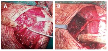
Figure 6 Intraoperative photos.
A: The headless Herbert screw is inserted and the larger fragments have been stabilized; B: The T-minus plate has been positioned acting as a buttress plate. The biceps tendon was found to be stable and was protected during the procedure.
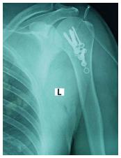
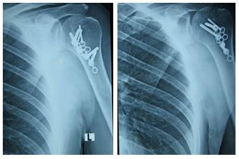
Figure 7 Immediate post-op X-ray of the left shoulder, showing excellent fracture reduction.
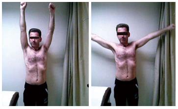
Figure 8 At the latest follow-up, the patient demonstrated pain-free, full range of movement of the left shoulder.
Figure 9 Eighteen months after surgery, X-ray examination of the left shoulder shows full union of fracture without hardware movement or failure.
- Citation: Nikolaou VS, Chytas D, Tyrpenou E, Babis GC. Two-level reconstruction of isolated fracture of the lesser tuberosity of the humerus. World J Clin Cases 2014; 2(6): 219-223
- URL: https://www.wjgnet.com/2307-8960/full/v2/i6/219.htm
- DOI: https://dx.doi.org/10.12998/wjcc.v2.i6.219


