Copyright
©2014 Baishideng Publishing Group Inc.
World J Clin Cases. Oct 16, 2014; 2(10): 596-603
Published online Oct 16, 2014. doi: 10.12998/wjcc.v2.i10.596
Published online Oct 16, 2014. doi: 10.12998/wjcc.v2.i10.596
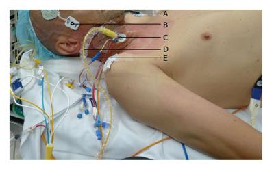
Figure 1 Monitoring used for redo-stenotomy and aortic valve replacement.
A: Cerebral oximeter (Invos, Somanetics®); B: Bispectral index; C: 9 French Internal jugular sheath; D: Continuous cardiac output and mixed venous oximetry measured with a fiberoptic pulmonary artery catheter (Edwards Lifesciences, Irvine CA); E: 4-lumen central venous catheter.
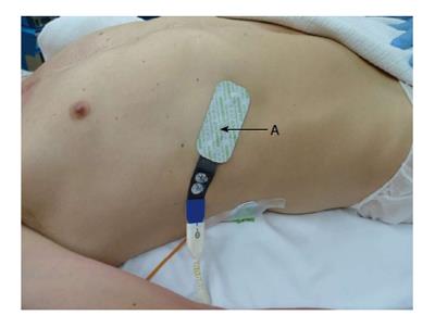
Figure 2 Hepatic tissue oxygenation measured by positioning an oximetry disposable transducer between the ribs and over the liver.
A: Hepatic oximeter (Invos, Somanetics).
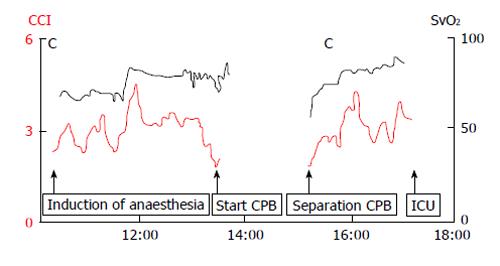
Figure 3 Intraoperative continuous cardiac index and mixed venous oxygenations tracings measured from the pulmonary artery catheter displayed on a Vigilance™ Monitor (Edwards Lifesciences, Irvine CA).
ICU: Intensive care unit; CPB: Cardiopulmonary bypass; CCI: Continuous cardiac index.
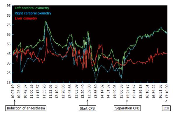
Figure 4 Cerebral and hepatic tissue oxygenation tracings measured with a cerebral/somatic oximeter (Invos Somanetics®) throughout the surgery and during cardiopulmonary bypass.
ICU: Intensive care unit; CPB: Cardiopulmonary bypass.
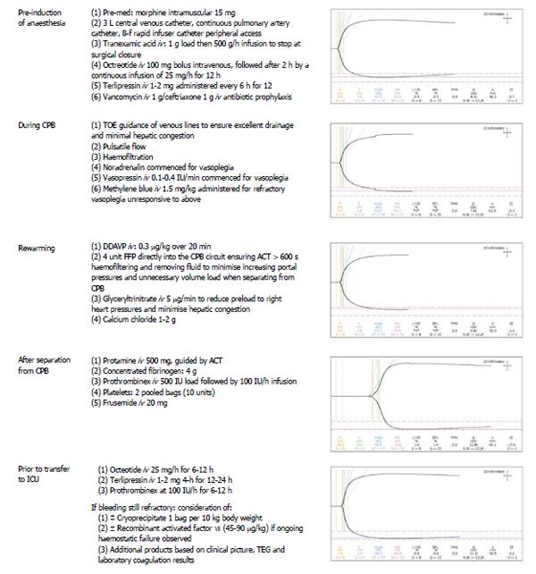
Figure 5 Perioperative thromboelastrography tracings observed in this case with corresponding haemostatic management action or planned strategy.
DDAVP: Desmopressin acetate; FFP: Fresh frozen plasma; CPB: Cardiopulmonary bypass; TEG: Thromboelastography; TOE: Transoesophageal echocardiography; IV: Intravenous; ACT: Activated clotting time.
- Citation: Weinberg L, Kearsey I, Tjoakarfa C, Matalanis G, Galvin S, Carson S, Bellomo R, McNicol L, McCall P. Haemostatic management for aortic valve replacement in a patient with advanced liver disease. World J Clin Cases 2014; 2(10): 596-603
- URL: https://www.wjgnet.com/2307-8960/full/v2/i10/596.htm
- DOI: https://dx.doi.org/10.12998/wjcc.v2.i10.596


