Copyright
©The Author(s) 2024.
World J Clin Cases. Apr 16, 2024; 12(11): 1990-1995
Published online Apr 16, 2024. doi: 10.12998/wjcc.v12.i11.1990
Published online Apr 16, 2024. doi: 10.12998/wjcc.v12.i11.1990
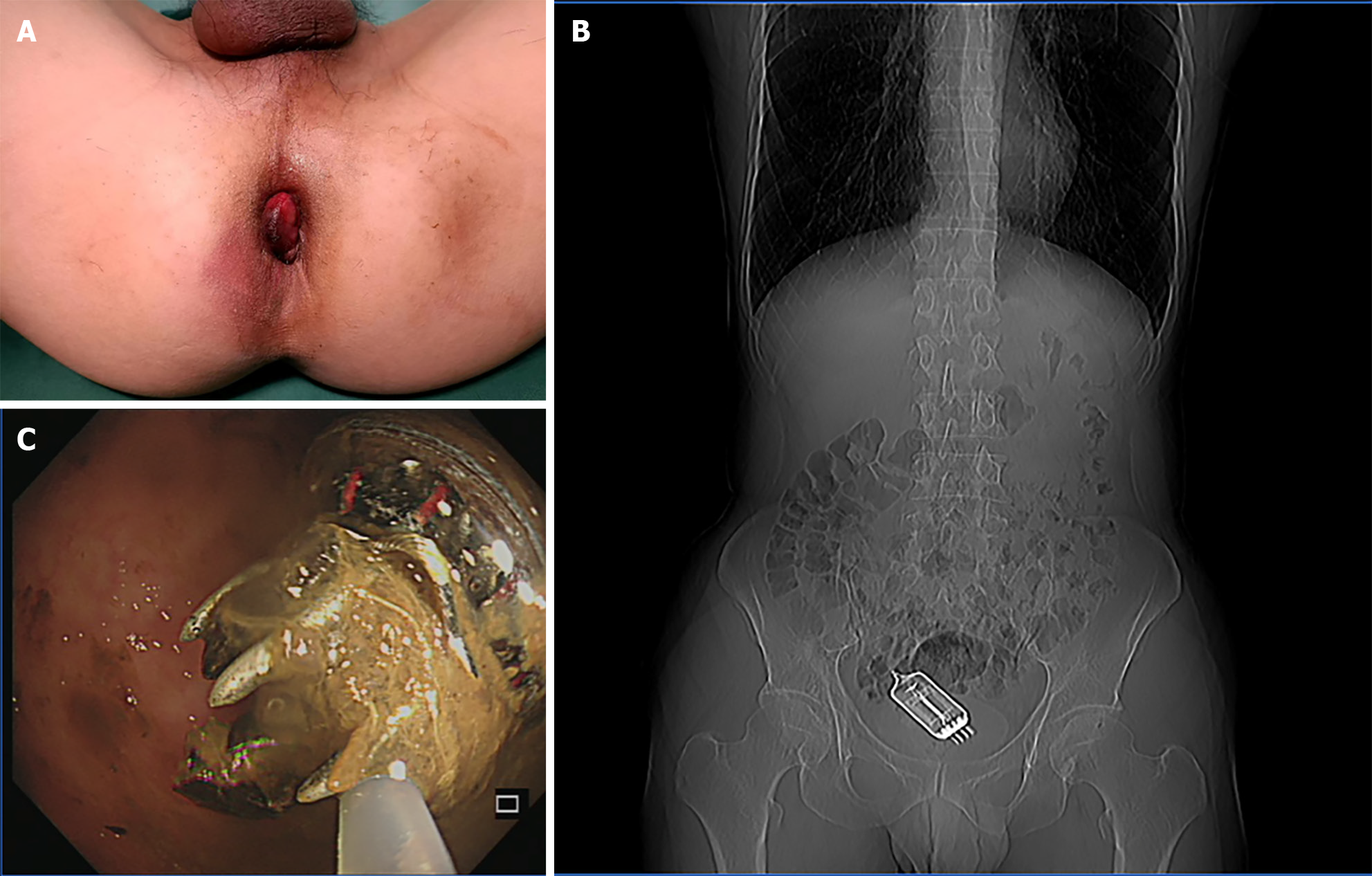
Figure 1 Computed tomography localization of the intestinal foreign body before surgery.
A: The patient had anal opening edema, acute prolapsed internal anal hemorrhoids, and surface congestion and erosion prior to surgery; B: Preoperatively, the foreign body was located at the junction of the rectum and sigmoid colon by computed tomography scanning; C: During preoperative colonoscopy, a foreign body was found at the junction of the rectum and sigmoid colon, with the tail end wrapped in intestinal secretions.
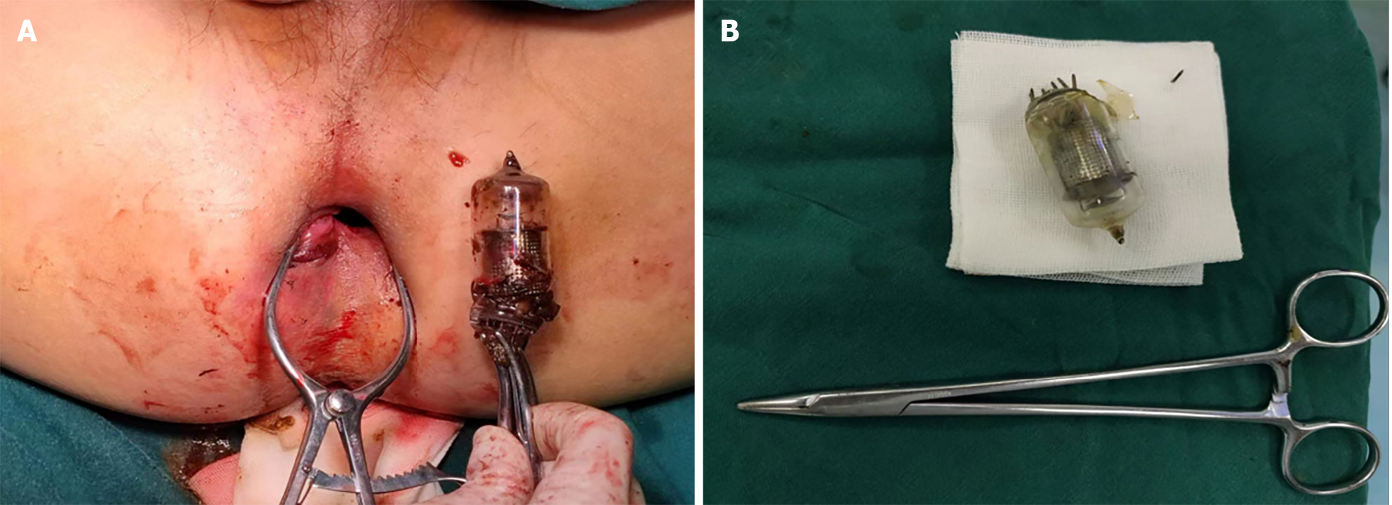
Figure 2 Postoperative foreign body morphology.
A: The foreign body was tractioned to the anal opening with the aid of digestive endoscopy, and was then removed by manipulation with the aid of a comb-type pulling tool; B: Comparison of foreign object to a needle receptacle.
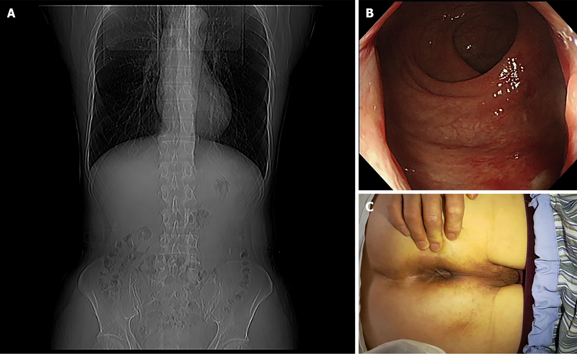
Figure 3 Postoperative evaluation of gastrointestinal foreign body and intestinal lumen status.
A: After 3 d of postoperative follow-up, no foreign objects or residues were found on computed tomography scanning; B: 3 d later, a follow-up colonoscopy was performed, and there was no damage to the intestinal cavity and no foreign body residue; C: 3 d after surgery, the symptoms of internal prolapsed hemorrhoids disappeared.
- Citation: Zhou PF, Lu JG, Zhang JD, Wang JW. Colonoscopy-assisted removal of an impaction foreign body at the rectosigmoid junction: A case report. World J Clin Cases 2024; 12(11): 1990-1995
- URL: https://www.wjgnet.com/2307-8960/full/v12/i11/1990.htm
- DOI: https://dx.doi.org/10.12998/wjcc.v12.i11.1990


