Copyright
©The Author(s) 2024.
World J Clin Cases. Apr 16, 2024; 12(11): 1980-1989
Published online Apr 16, 2024. doi: 10.12998/wjcc.v12.i11.1980
Published online Apr 16, 2024. doi: 10.12998/wjcc.v12.i11.1980
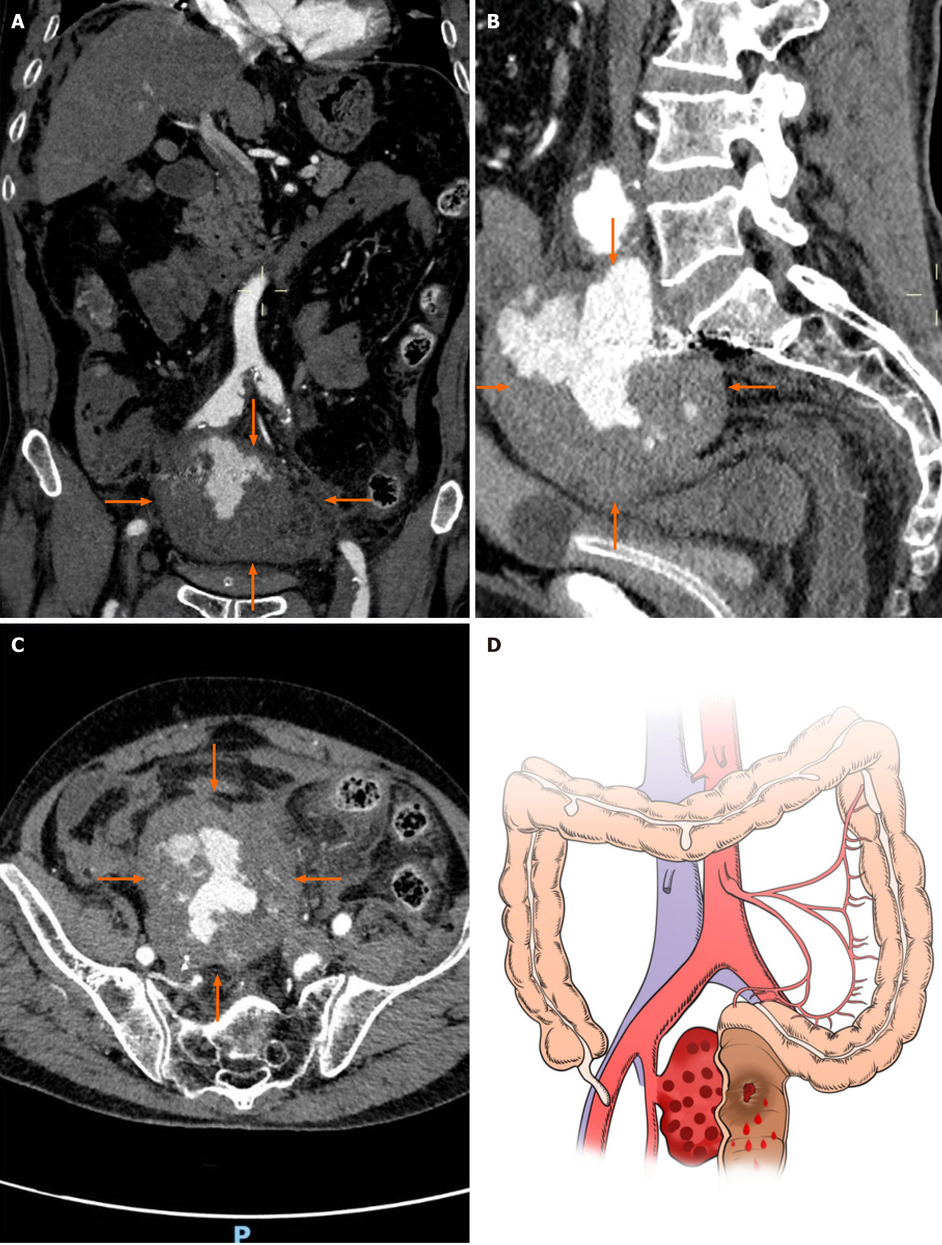
Figure 1 Computed tomography images of the abdomen and pelvis.
A: Coronal computed tomography (CT) image showing an oval-shaped mass with mixed high and low densities near the bifurcation of the right internal iliac artery (orange arrow); B: Coronal CT image showing an internal iliac aneurysm occupying the intrapelvic space, with a vertical diameter of approximately 7 centimeters, and suspected to be communicating with the rectum; C: Axial CT image showing that the internal iliac artery aneurysm had transverse and anteroposterior diameters of approximately 9.5 cm and 8.5 cm, respectively, suggesting the possibility of aneurysm rupture and hematoma formation; D: The illustration shows that the rectal wall was invaded by the right internal iliac artery aneurysm, which led to rectal bleeding.
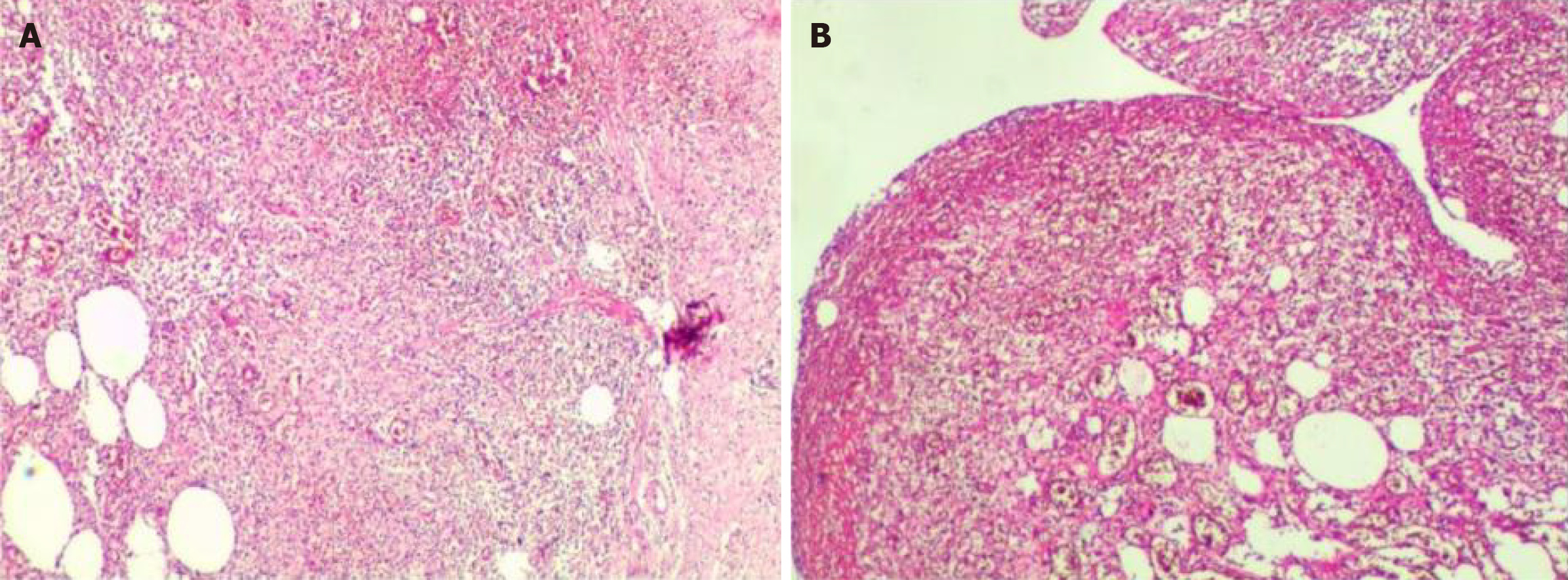
Figure 2 Pathological diagnosis (magnification 10 × 10).
A: Pathological examination of the aneurysm wall revealed hemorrhage and fibrous exudation; B: Rectal pathologic examination showed ulceration with perforation, mucosal edema, submucosal and serosal purulent inflammation with bleeding and necrosis, consistent with changes related to intestinal perforation. Ulceration was noted at one end of the rectum, while the other end was free of disease. Additionally, reactive hyperplasia of perienteric lymph nodes (17 in total) was observed.
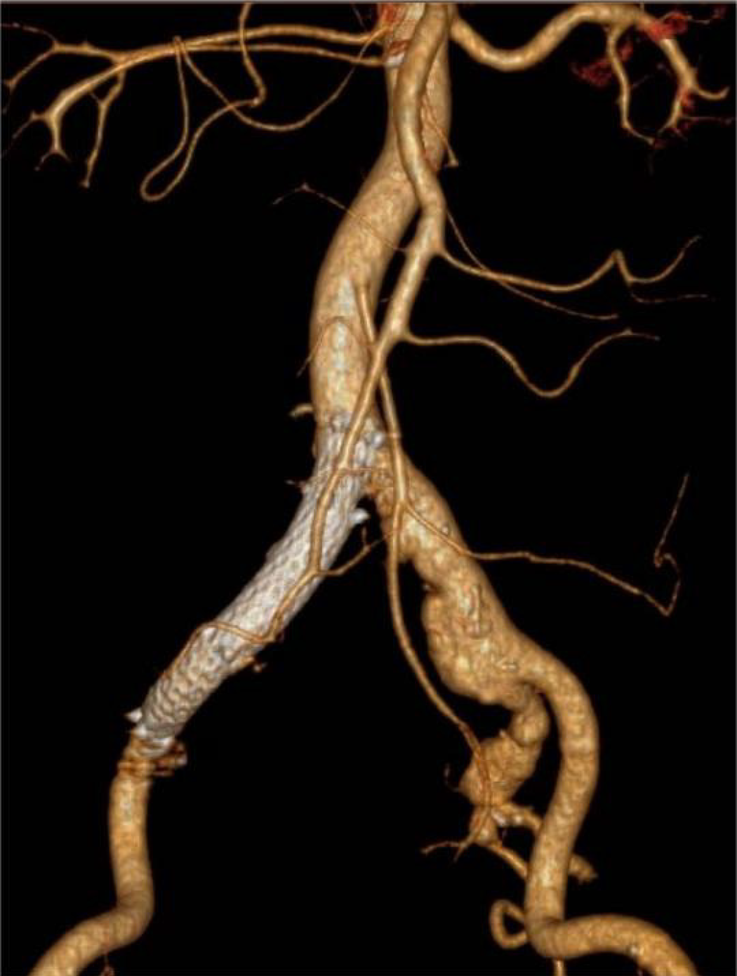
Figure 3 Computed tomography angiography images showing the stent placement site.
Postoperative computed tomography angiography suggested that the covered stent deployed in the iliac artery was fixed in its predetermined location and that the pelvic hematoma had decreased in size when compared to its previous size.
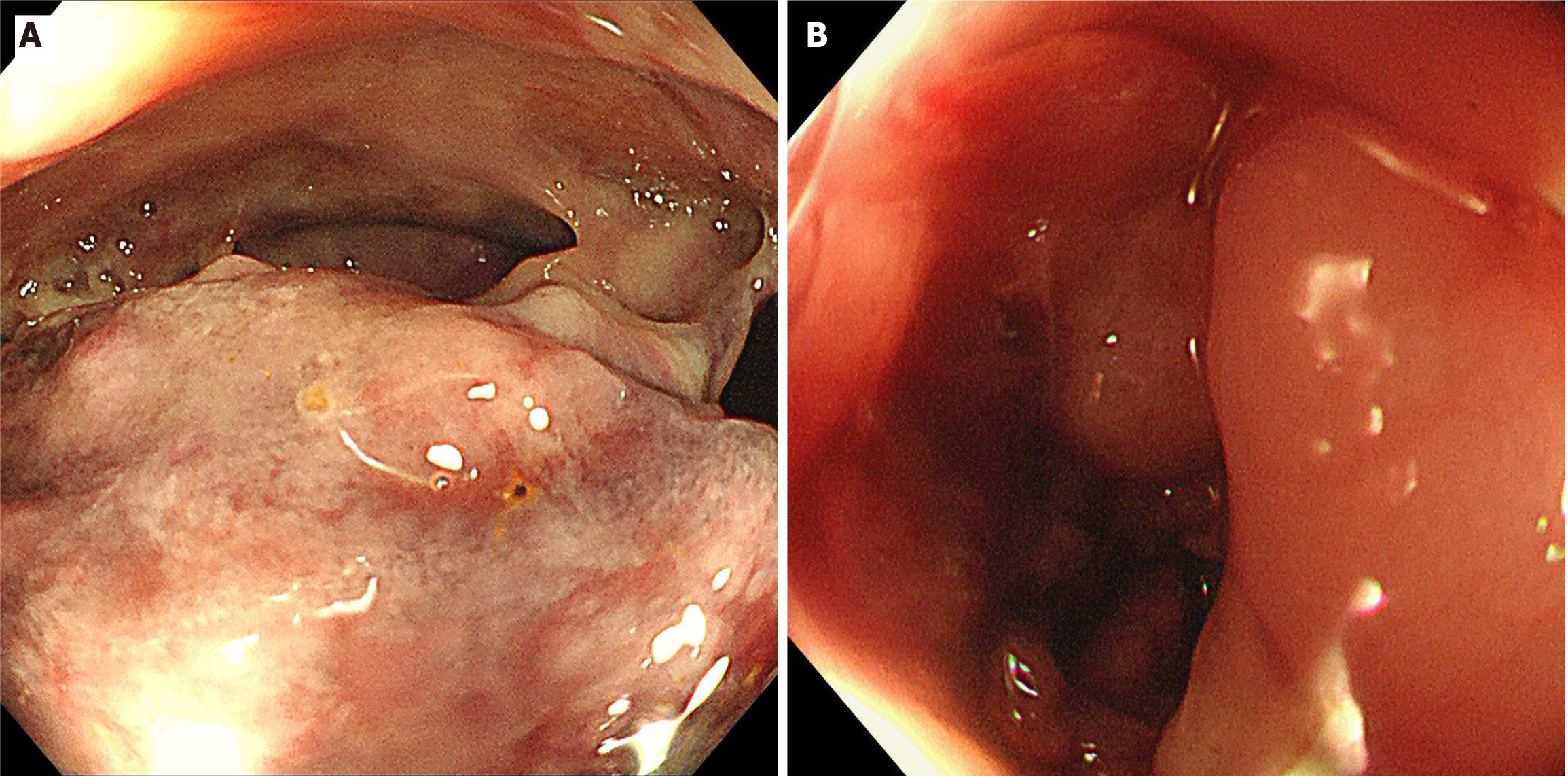
Figure 4 Colonoscopic images.
A: Preoperative colonoscopy revealed a large neoplasm invading the pelvic cavity approximately 16 cm from the anus and covered with a turbid coating, blood clots, and purulent secretions, with bubbles visible on the surface, suggesting the possibility of a rectal neoplasm with perforation; B: Intraoperative colonoscopy revealed a perforation 0.8 cm in the rectum located 15 cm from the anus and surrounded by neoplastic growth and purulent secretions.
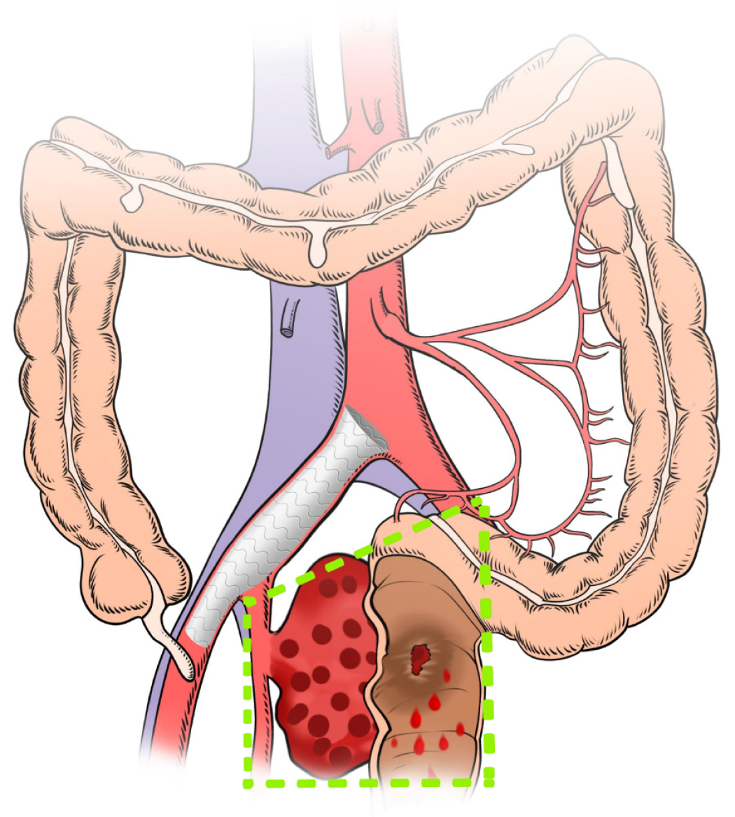
Figure 5 A hand-drawn drawing of the scope of the surgical resection.
Illustration showing the extent of surgical excision: Hematoma evacuation, aneurysm resection, resection of the diseased part of the rectum, intestinal anastomosis, and prophylactic ileostomy (as shown within the area marked by the green line).
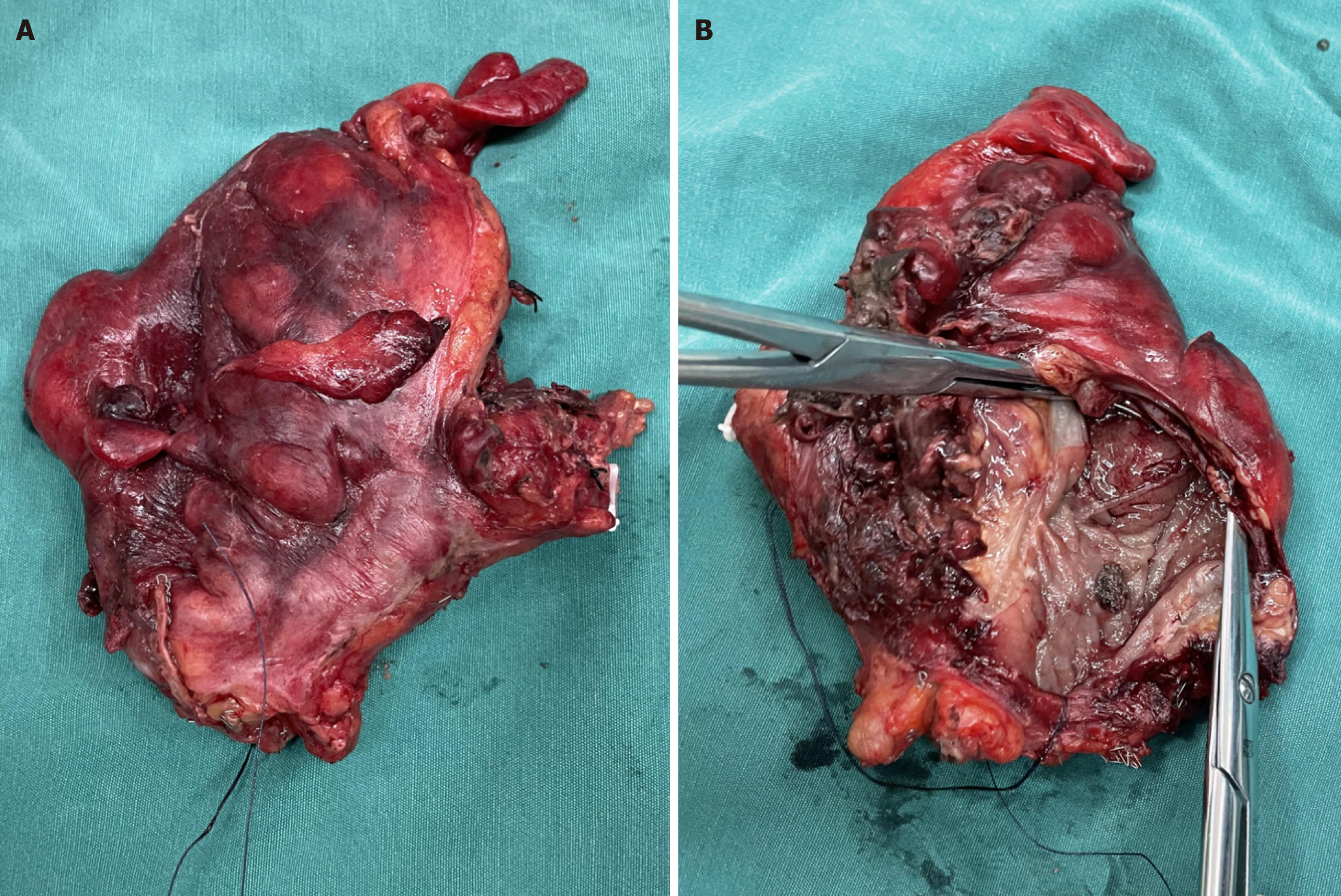
Figure 6 Photograph of the resected rectum.
A: Macroscopic appearance of the rectal surgery specimen; B: The appearance of the luminal cross-section of the incised rectal wall shows that on one side of the intestinal wall, there was visible congestion, erosion, and edematous tissue, with purulent necrotic secretions attached, and a perforation approximately 0.8 cm in size in the intestinal wall.
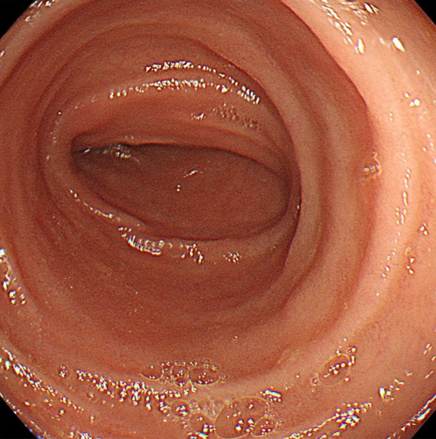
Figure 7 Postoperative colonoscopic images.
Six months after discharge, colonoscopy revealed a normal anastomotic site, with no intestinal stenosis, ulceration, or mass formation.
- Citation: Li F, Zhao B, Liu YQ, Chen GQ, Qu RF, Xu C, Long Z, Wu JS, Xiong M, Liu WH, Zhu L, Feng XL, Zhang L. Hematochezia due to rectal invasion by an internal iliac artery aneurysm: A case report. World J Clin Cases 2024; 12(11): 1980-1989
- URL: https://www.wjgnet.com/2307-8960/full/v12/i11/1980.htm
- DOI: https://dx.doi.org/10.12998/wjcc.v12.i11.1980


