Copyright
©The Author(s) 2022.
World J Clin Cases. Nov 6, 2022; 10(31): 11508-11516
Published online Nov 6, 2022. doi: 10.12998/wjcc.v10.i31.11508
Published online Nov 6, 2022. doi: 10.12998/wjcc.v10.i31.11508
Figure 1 Timeline.
PCR: Polymerase chain reaction; MI: Myocardial infarction; RCA: Right coronary artery; IVUS: Intravascular ultrasound; OCT: Optical coherence tomography; RFR: Resting full-cycle ratio; COVID-19: Coronavirus disease 2019.
Figure 2 Emergency coronary angiogram: Left coronary artery.
Angiography reveals no significant stenoses. A: Left coronary artery (LCA) in the conventional right anterior oblique projection; B: LCA in the caudal right anterior oblique projection; C: LCA in the cranial posteroanterior projection; D: LCA in the cranial left anterior oblique projection.
Figure 3 Emergency coronary angiogram: Right coronary artery and left ventriculography.
The images show intraluminal filling defects (red arrows) starting in the middle third of the right coronary artery (RCA) and extending to the distal third, affecting the posterior descending and posterolateral RCA branches. A: RCA in the conventional left anterior oblique projection; B: RCA in the cranial left anterior oblique projection; C: Left ventriculography (LV) in systole demonstrates inferior akinesia (yellow arrows); D: LV in diastole.
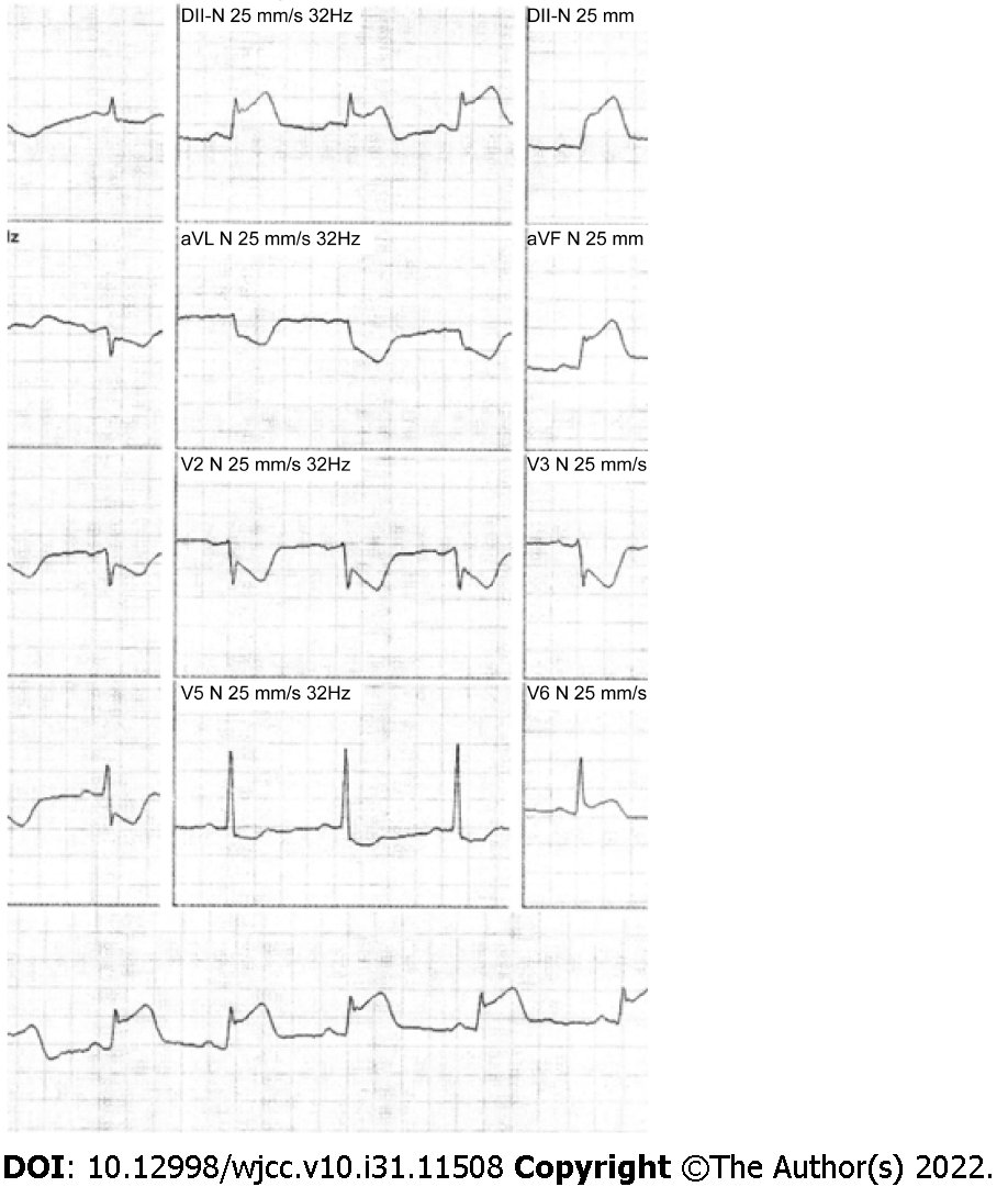
Figure 4 Electrocardiogram on admission.
The results show sinus rhythm with ST-segment elevation in the inferior wall and reciprocal changes in the anterolateral leads.
Figure 5 Intravascular ultrasound images from two angles.
A: Right coronary artery (RCA) in the conventional left anterior oblique projection; B: RCA in the cranial left anterior oblique projection; C: An incipient plaque; D: A concentric plaque-causing moderate luminal stenosis; E-H: A subacute-appearing thrombus with a layered light to dark-gray appearance with white patches and less clear delineations. Moderate-to-severe signal attenuation may also be observed, likely due to the deviation or absorption of ultrasound waves by the subacute thrombus; I: An acute thrombus (asterisk) with a bright appearance, clear outline, and no signal attenuation; J: Discreet intimal thickening.
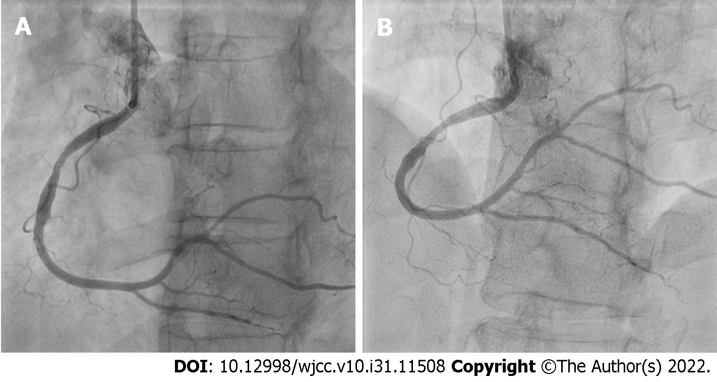
Figure 6 Reexamination of the right coronary artery after 7 d.
The images show nearly complete resolution of the right coronary artery (RCA) thrombus. A: RCA in the conventional left anterior oblique projection; B: RCA in the cranial left anterior oblique projection.
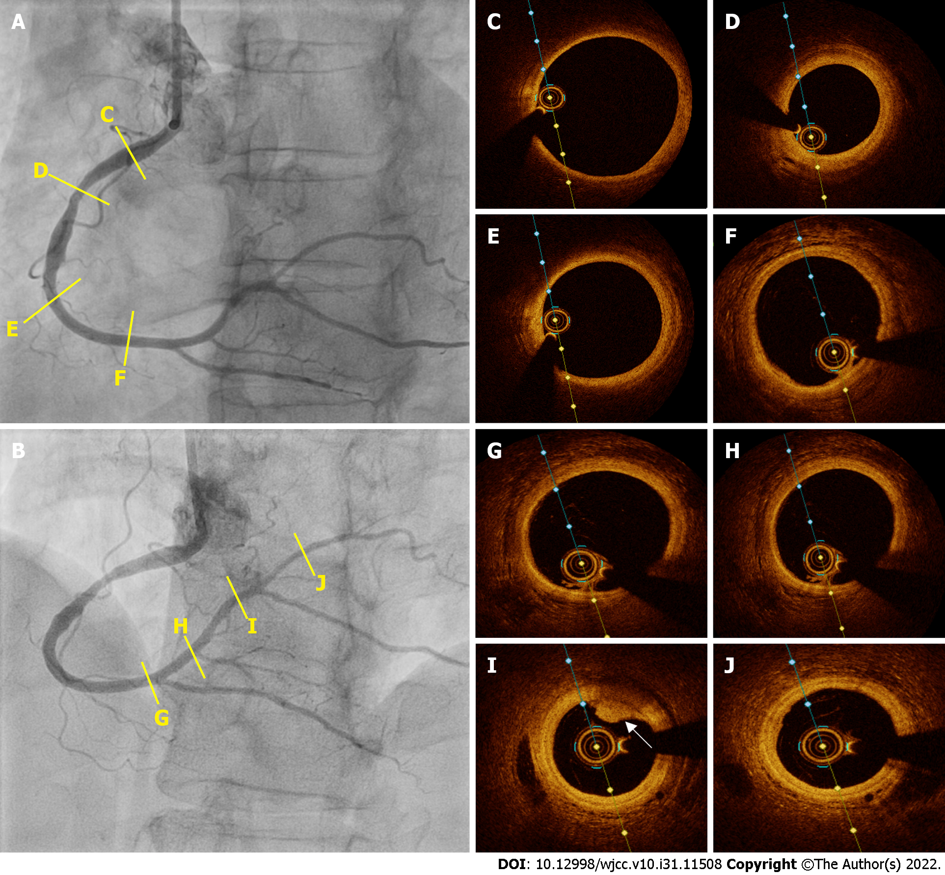
Figure 7 Optical coherence tomography images from two angles.
A: Right coronary artery (RCA) in the conventional left anterior oblique projection; B: RCA in the cranial left anterior oblique projection; C: A normal vessel with a three-layered structure; D: A fibrotic plaque; E and F: Normal vessels with a three-layer structure; G, H, and J: Normal vessels with a three-layer structure; I: Mild signal attenuation and easily delineated borders suggest white thrombus remnants (white arrow).
- Citation: Dall’Orto CC, Lopes RPF, Cancela MT, de Sales Padilha C, Pinto Filho GV, da Silva MR. Extensive right coronary artery thrombosis in a patient with COVID-19: A case report. World J Clin Cases 2022; 10(31): 11508-11516
- URL: https://www.wjgnet.com/2307-8960/full/v10/i31/11508.htm
- DOI: https://dx.doi.org/10.12998/wjcc.v10.i31.11508


