Copyright
©The Author(s) 2022.
World J Clin Cases. Jul 16, 2022; 10(20): 7037-7044
Published online Jul 16, 2022. doi: 10.12998/wjcc.v10.i20.7037
Published online Jul 16, 2022. doi: 10.12998/wjcc.v10.i20.7037
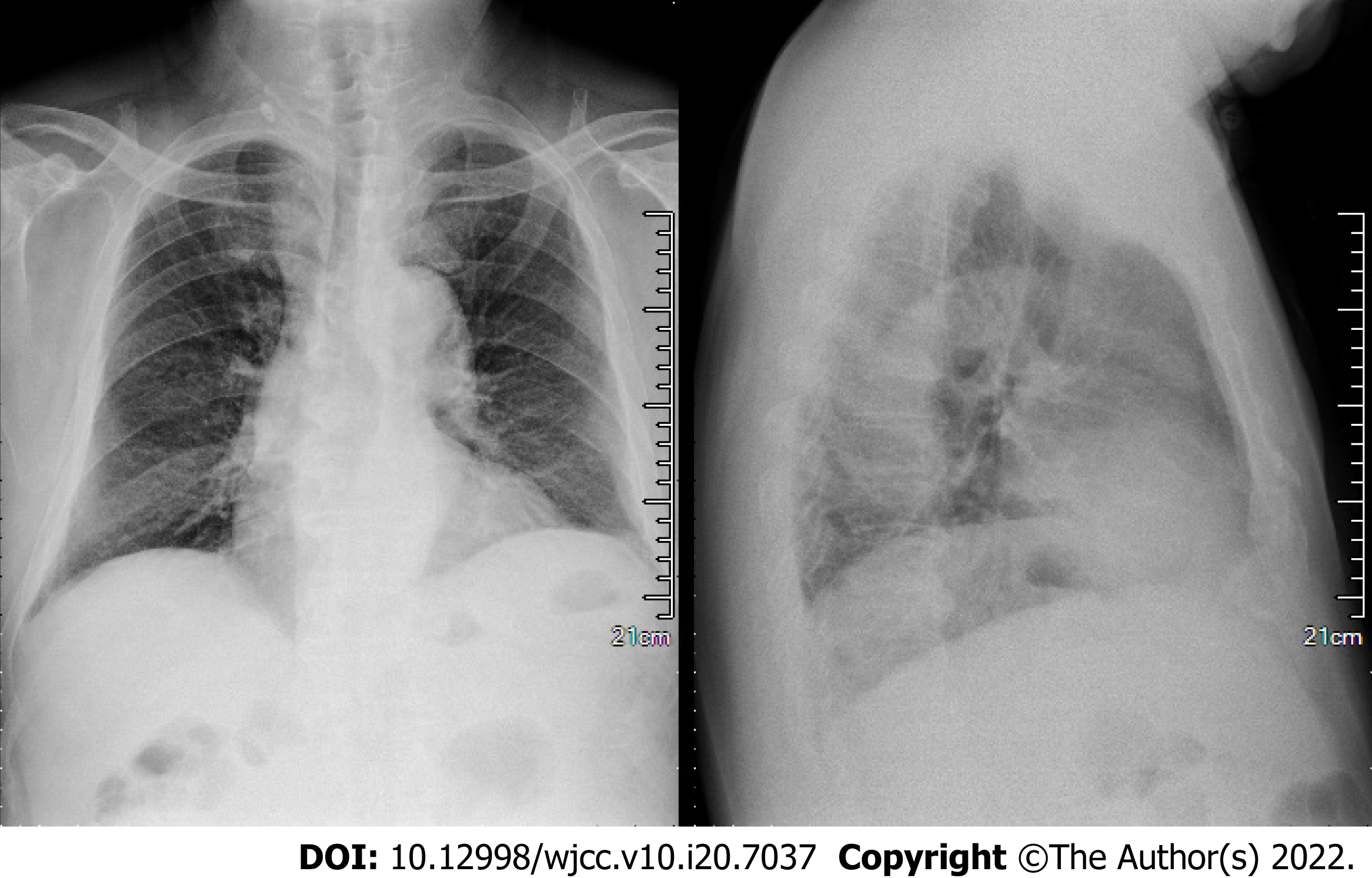
Figure 1 Chest X-ray.
No significant cardiopulmonary abnormalities were seen.
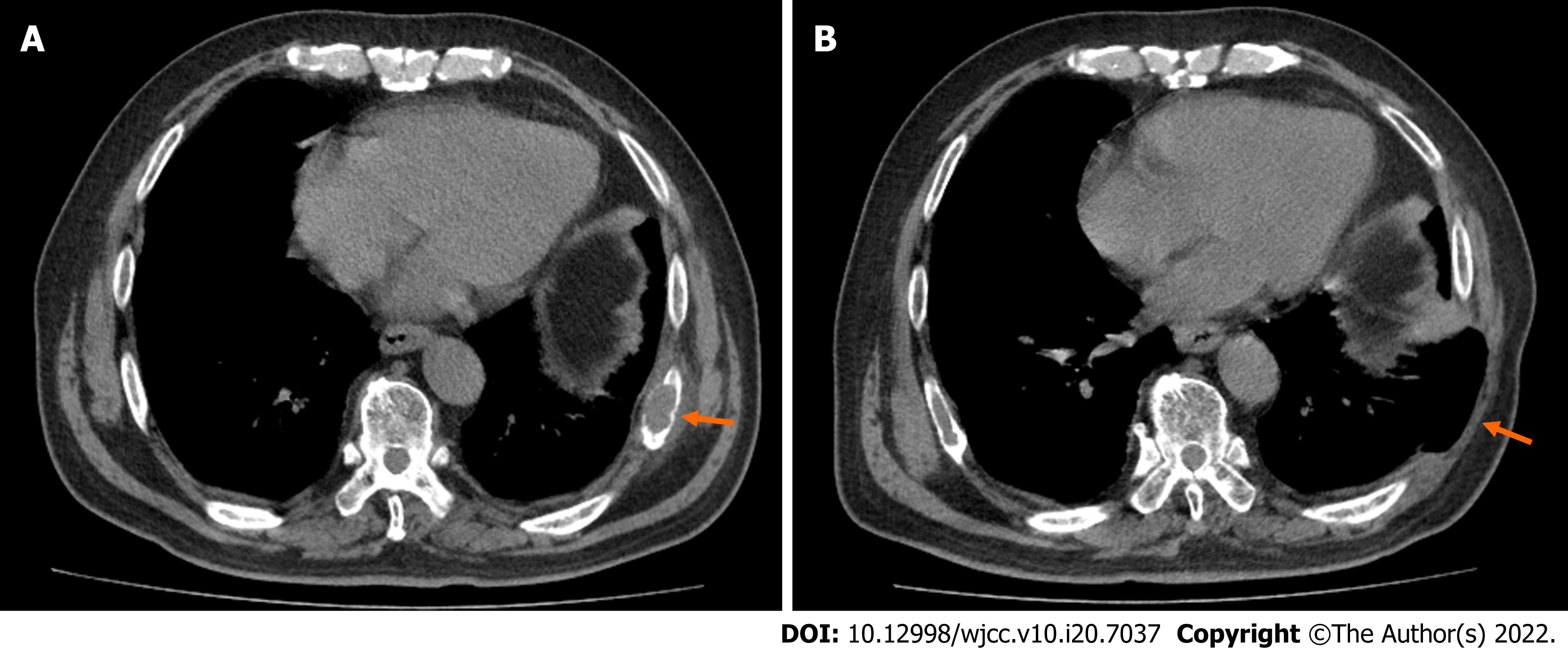
Figure 2 Comparison of computed tomography (CT) images of left eighth rib mass before and after surgery.
A: CT scan of lung before surgery showed that the left eighth rib mass was accompanied by bone destruction (orange arrow); B: Lung CT scan after surgery showed changes after resection of the left eighth rib (orange arrow).
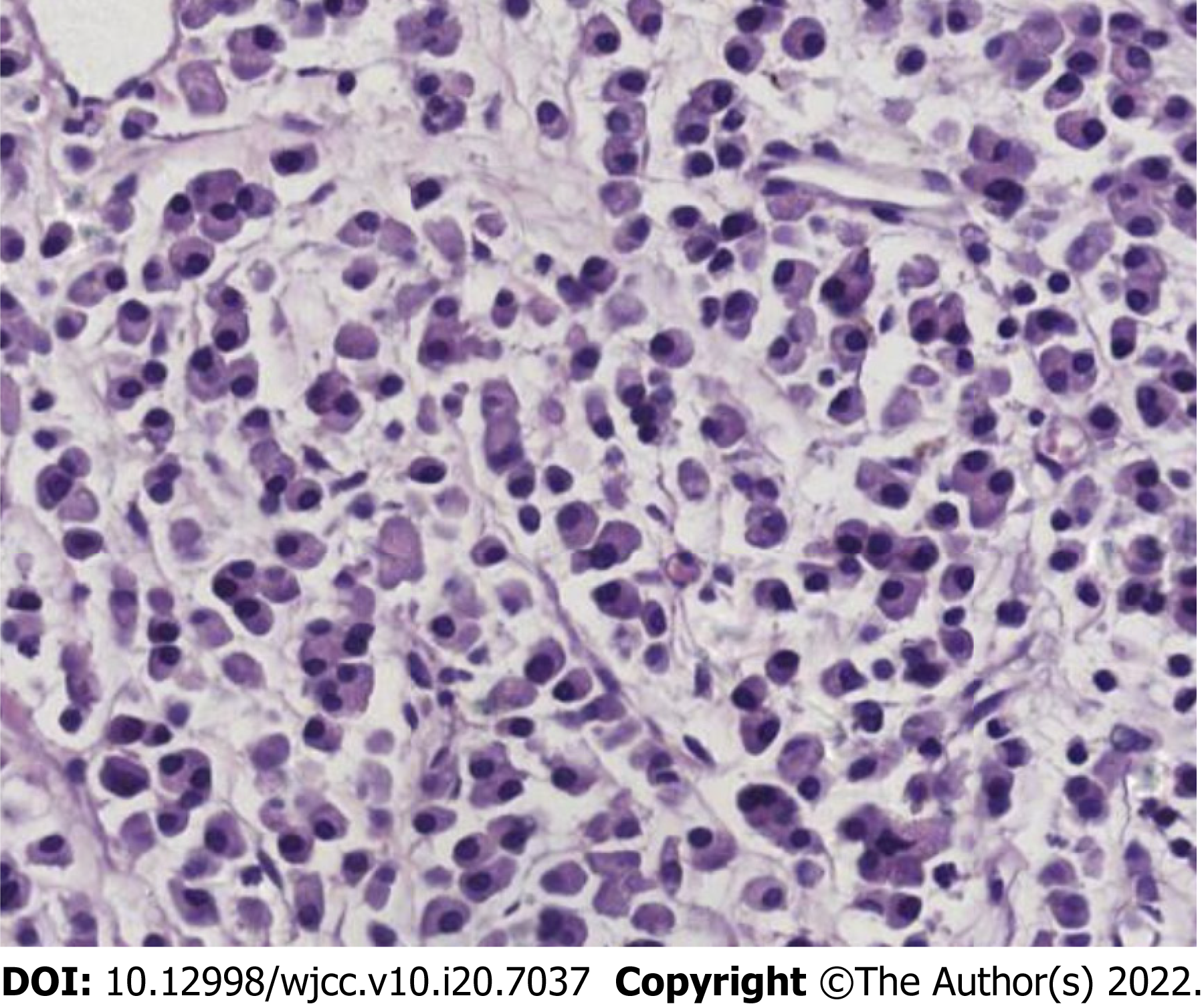
Figure 3 Postoperative pathology showed that a large number of immature plasma cells proliferated after decalcification (HE, × 400).
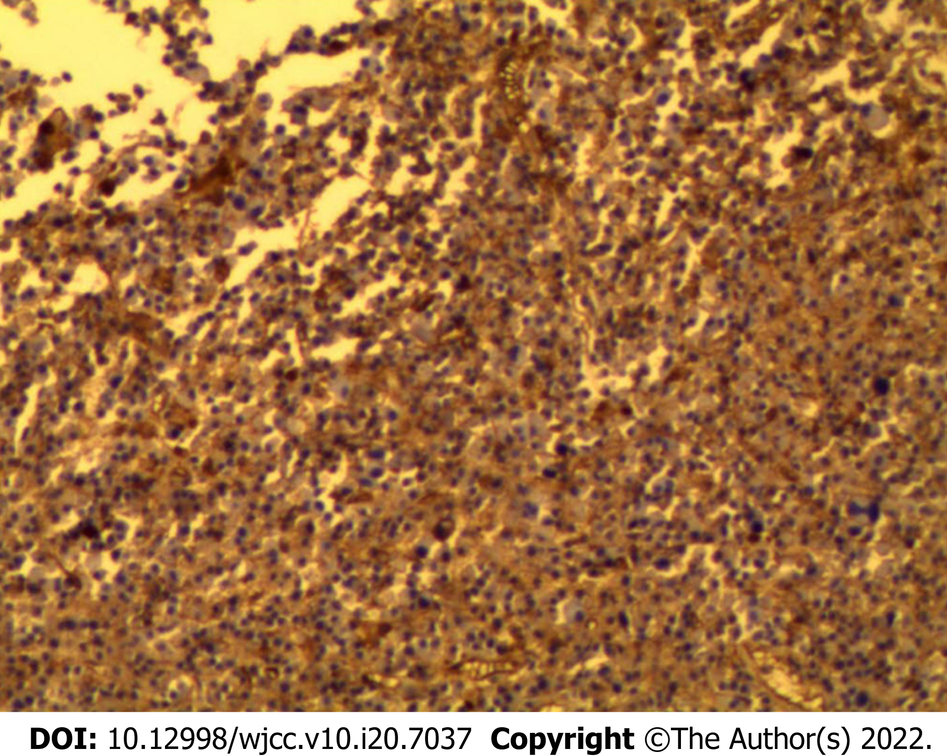
Figure 4 Immunohistochemical staining showed diffuse CD 138(+) in the tumor cell membrane (× 100).
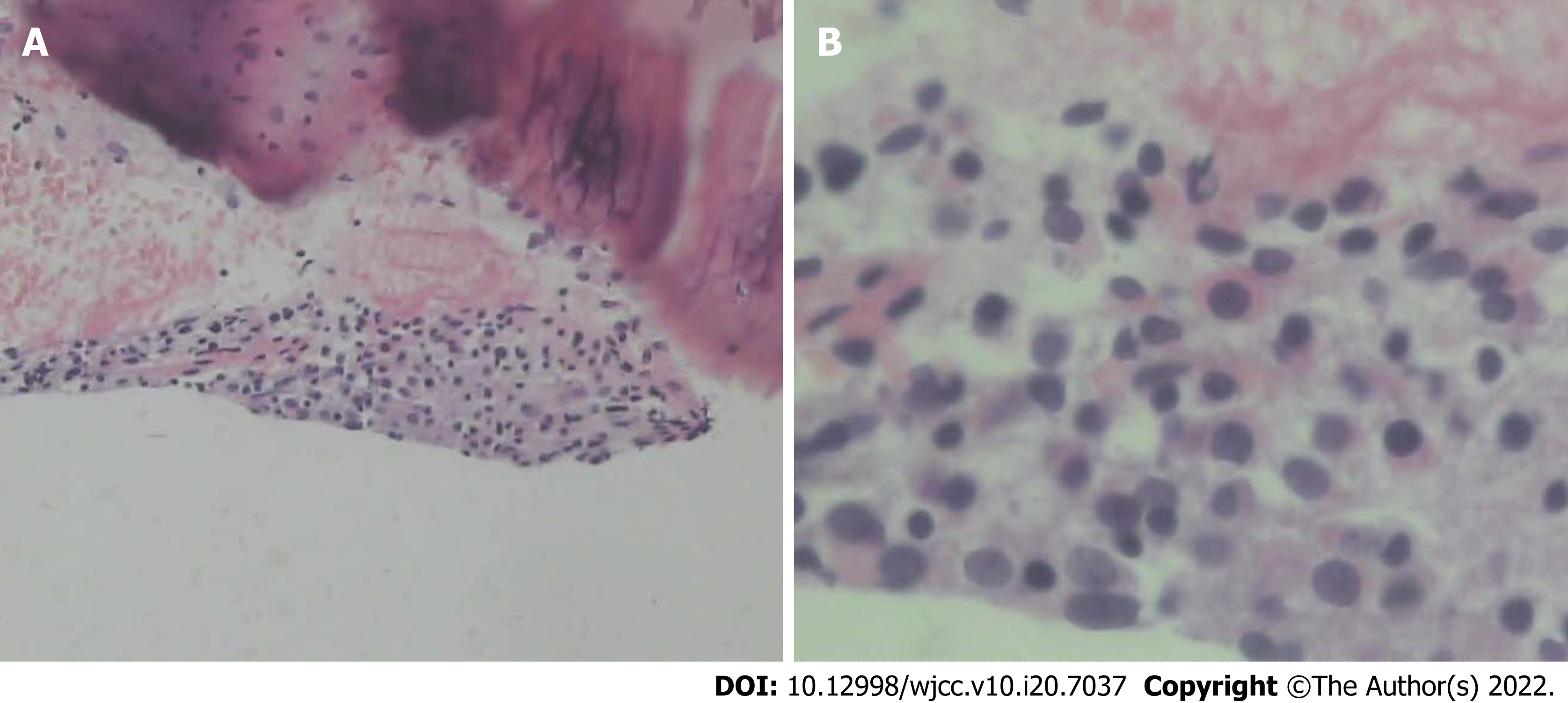
Figure 5 Pathology of rib biopsy.
A: The pathological findings of rib biopsy showed that it was mainly striated muscle tissue, with a small amount of connective tissue, skin and bone tissue, and short spindle cells (HE, × 100); B: Rib biopsy pathology showed that under high magnification, some cells were short spindle-shaped, and the nuclei were slightly dark stained, with non-nuclear divisions (HE, × 400).
- Citation: Yao J, He X, Wang CY, Hao L, Tan LL, Shen CJ, Hou MX. Solitary plasmacytoma of the left rib misdiagnosed as angina pectoris: A case report . World J Clin Cases 2022; 10(20): 7037-7044
- URL: https://www.wjgnet.com/2307-8960/full/v10/i20/7037.htm
- DOI: https://dx.doi.org/10.12998/wjcc.v10.i20.7037


