Copyright
©The Author(s) 2022.
World J Clin Cases. Jul 16, 2022; 10(20): 7006-7012
Published online Jul 16, 2022. doi: 10.12998/wjcc.v10.i20.7006
Published online Jul 16, 2022. doi: 10.12998/wjcc.v10.i20.7006
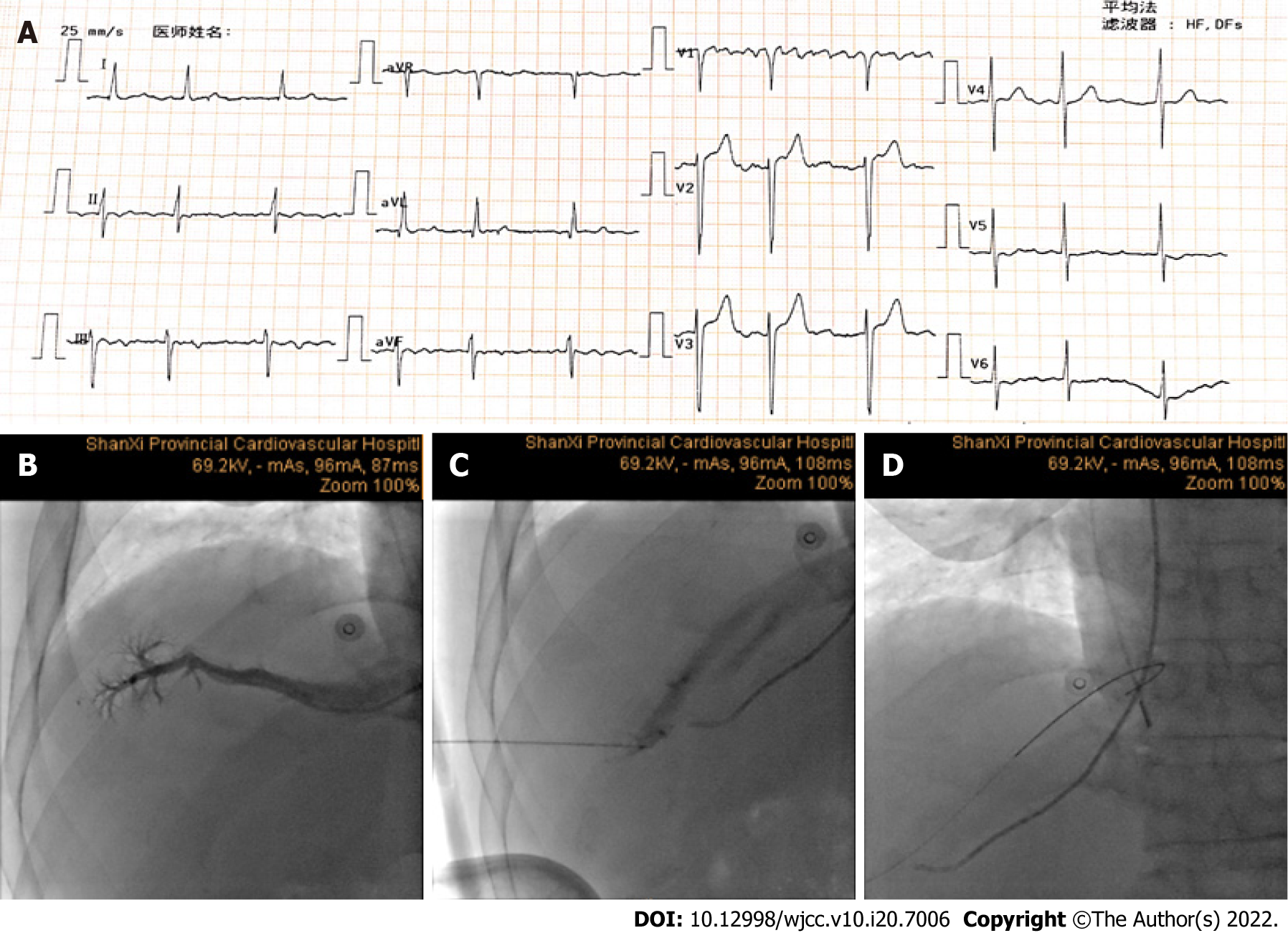
Figure 1 Serial fluoroscopic still frames during the procedure.
A: Electrocardiogram before the operation; B: JR4.0 catheter was inserted into the hepatic vein via the superior vena cava and then a contrast agent was injected to determine the main direction of the hepatic vein; C: Hepatic access was obtained under the guidance of angiography; D: A 0.035-inch Bentson wire was placed through the needle into the right atrium.
Figure 2 Visualization of the interatrial septal puncture.
A: Interatrial septal puncture under the guidance of intracardiac echocardiography (ICE), and the procedure presented a tent-like shape visualized by an orange line; B: ICE image: The ablation catheter in the left atrium, single access was obtained for the ablation; C: Anatomy of the heart at the right anterior view and the right view.
Figure 3 Electroanatomic map obtained during procedure.
A: Hepatic signal position is visualized by a green color; B: Left atrial electroanatomic map and pulmonary vein isolation radiofrequency ablation lesions; C: Electrocardiogram after the operation.
- Citation: Wang HX, Li N, An J, Han XB. Percutaneous transhepatic access for catheter ablation of a patient with heterotaxy syndrome complicated with atrial fibrillation: A case report. World J Clin Cases 2022; 10(20): 7006-7012
- URL: https://www.wjgnet.com/2307-8960/full/v10/i20/7006.htm
- DOI: https://dx.doi.org/10.12998/wjcc.v10.i20.7006


