Copyright
©The Author(s) 2022.
World J Clin Cases. Jul 16, 2022; 10(20): 6966-6973
Published online Jul 16, 2022. doi: 10.12998/wjcc.v10.i20.6966
Published online Jul 16, 2022. doi: 10.12998/wjcc.v10.i20.6966
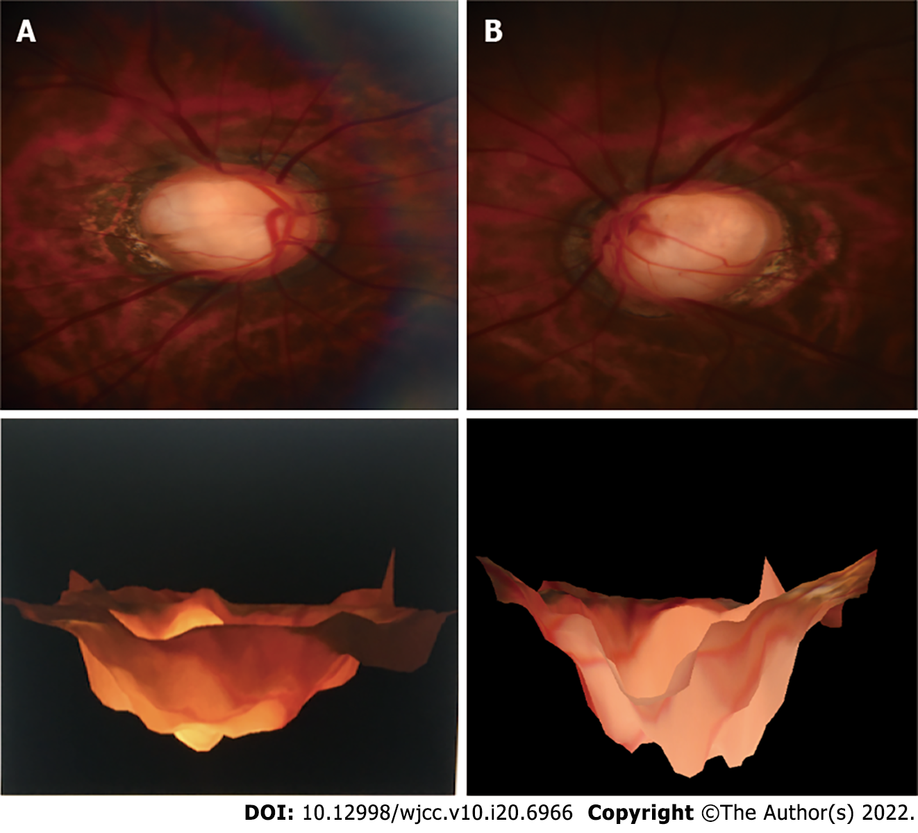
Figure 1 Stereoscopic color photography of the optic disc of the right (A) and left (B) eyes.
Figure 2 Three-dimensional optical coherence tomography optic disc mode of the optic disc of the right (A) and left (B) eyes.
Figure 3 En-face-optical coherence tomography and B-scan macular pattern of the macula of right (A) and left (B) eyes.
Figure 4 Angio-optical coherence tomography macular 6 mm × 6 mm pattern of the right (A) and left (B) eyes.
Figure 5 Optical coherence tomography B-scan macular pattern of right (A) and left (B) eye for 2 years after surgery.
Figure 6 Macular retinoschisis associated with optic disc coloboma may be the result of the combined force.
- Citation: Zhang W, Peng XY. Optic disc coloboma associated with macular retinoschisis: A case report. World J Clin Cases 2022; 10(20): 6966-6973
- URL: https://www.wjgnet.com/2307-8960/full/v10/i20/6966.htm
- DOI: https://dx.doi.org/10.12998/wjcc.v10.i20.6966


