Copyright
©The Author(s) 2022.
World J Clin Cases. Jul 6, 2022; 10(19): 6736-6743
Published online Jul 6, 2022. doi: 10.12998/wjcc.v10.i19.6736
Published online Jul 6, 2022. doi: 10.12998/wjcc.v10.i19.6736
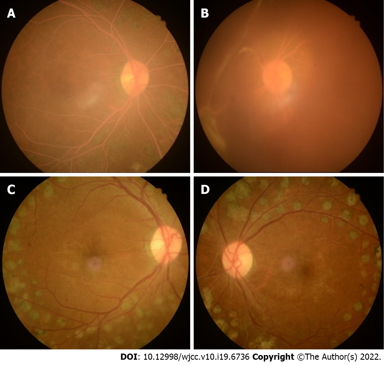
Figure 1 Fundus images with lipemia retinalis and the fundus in the same case with normal blood lipids.
A, B: The right and left eye fundus images with lipemia retinalis, respectively, and showed a pink–white color of the fundus, arteries, and veins. It had simultaneous vitreous hemorrhage (B); C, D: The right and left eye fundus images in the same case with normal blood lipids.
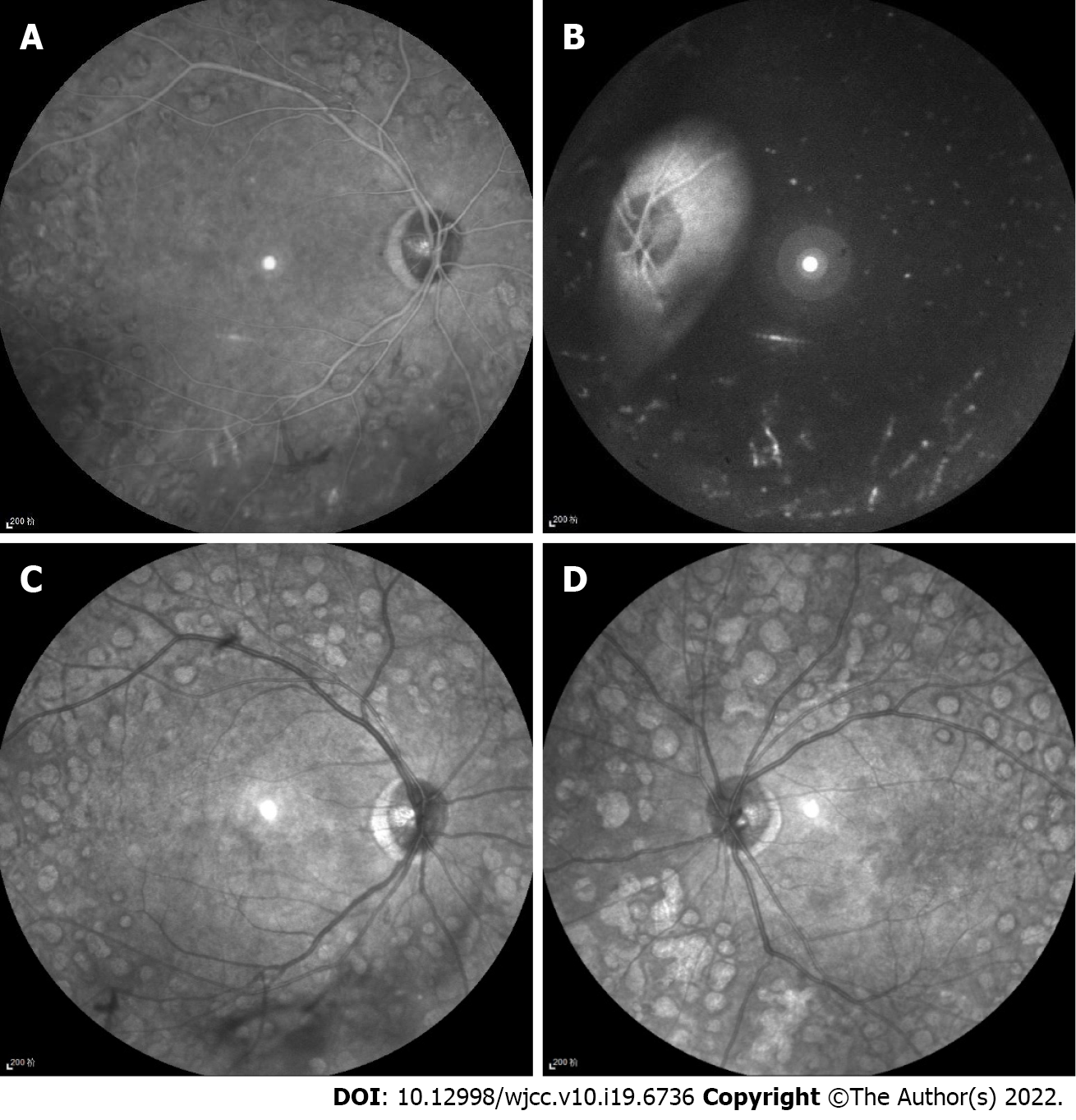
Figure 2 Infrared images with lipemia retinalis and infrared images of the same case with normal blood lipids.
A, B: Infrared images of lipemia retinalis showed hyperinfrared reflection of retinal vessels; C, D: Infrared images of the same case with normal blood lipids showed hypoinfrared reflection of retinal vessels.
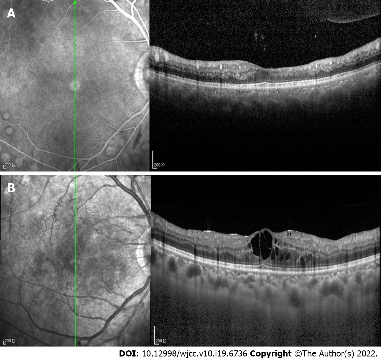
Figure 3 Optical coherence tomography with lipemia retinalis and optical coherence tomography of the same case with normal blood lipids.
A: The right eye Optical coherence tomography (OCT) with lipemia retinalis, which showed point-like high reflections in the retina, corresponding to the cross-section of retinal blood vessels, and medium reflections in the choroid big vessels; B: The right eye OCT of the same case with normal blood lipids.
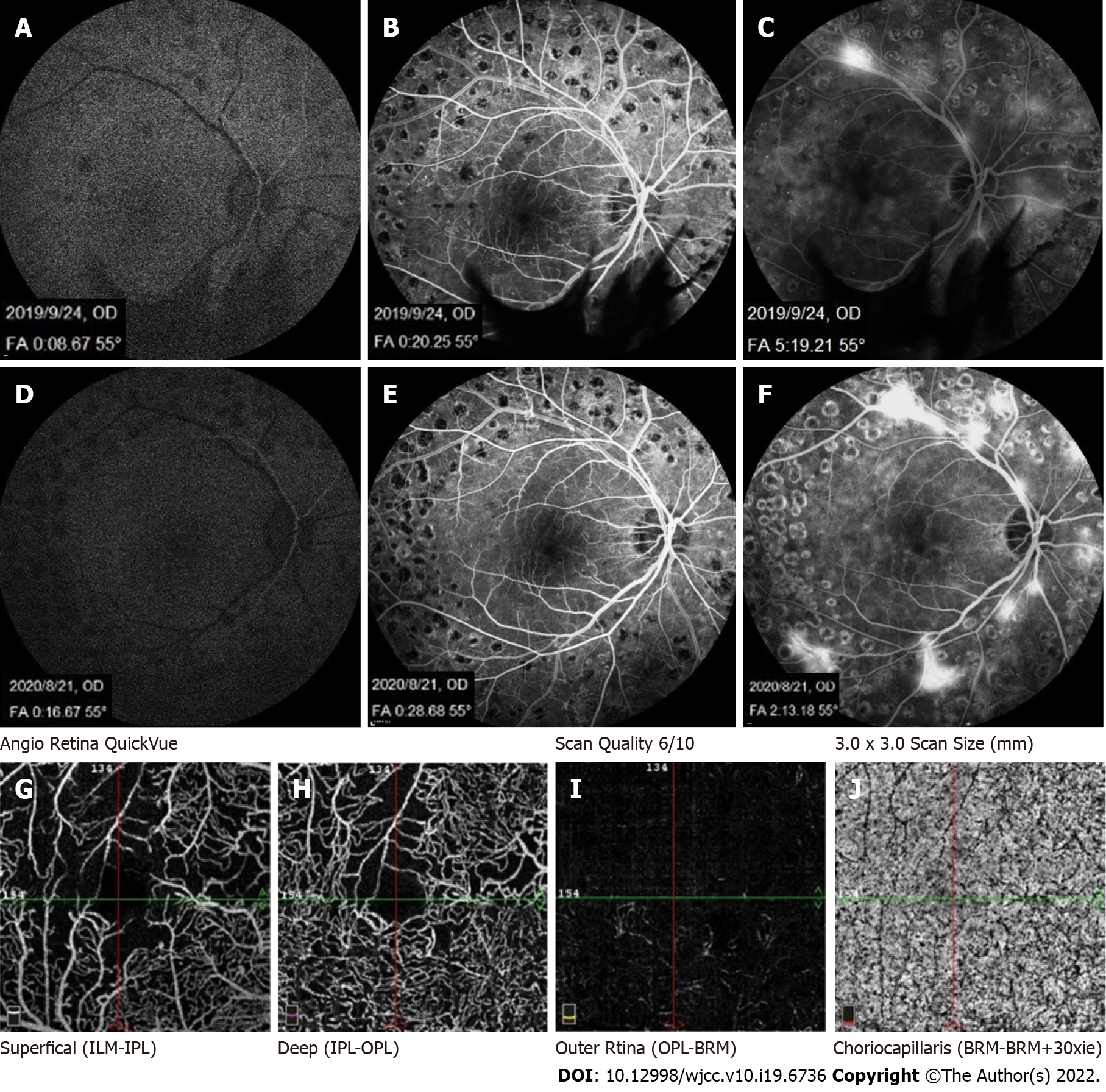
Figure 4 Fundus fluorescein angiography with lipemia retinalis, fundus fluorescein angiography of the present case with normal blood lipids, and optical coherence tomography angiography of this case with lipemia retinalis.
A-C: The fundus fluorescein angiography of this case with normal blood lipids; D, E, and F: The fundus fluorescein angiography of this case with lipemia retinalis (LR); A–F showed no significant difference in retinal filling time and fundus fluorescence between the patient’s hypertriglyceridemia condition and a normal blood lipid condition; G-J: The optical coherence tomography angiography of LR, showing a nonperfusion area consistent with fundus fluorescein angiography.
- Citation: Zhang SJ, Yan ZY, Yuan LF, Wang YH, Wang LF. Multimodal imaging study of lipemia retinalis with diabetic retinopathy: A case report. World J Clin Cases 2022; 10(19): 6736-6743
- URL: https://www.wjgnet.com/2307-8960/full/v10/i19/6736.htm
- DOI: https://dx.doi.org/10.12998/wjcc.v10.i19.6736


