Copyright
©The Author(s) 2022.
World J Clin Cases. Jul 6, 2022; 10(19): 6548-6554
Published online Jul 6, 2022. doi: 10.12998/wjcc.v10.i19.6548
Published online Jul 6, 2022. doi: 10.12998/wjcc.v10.i19.6548
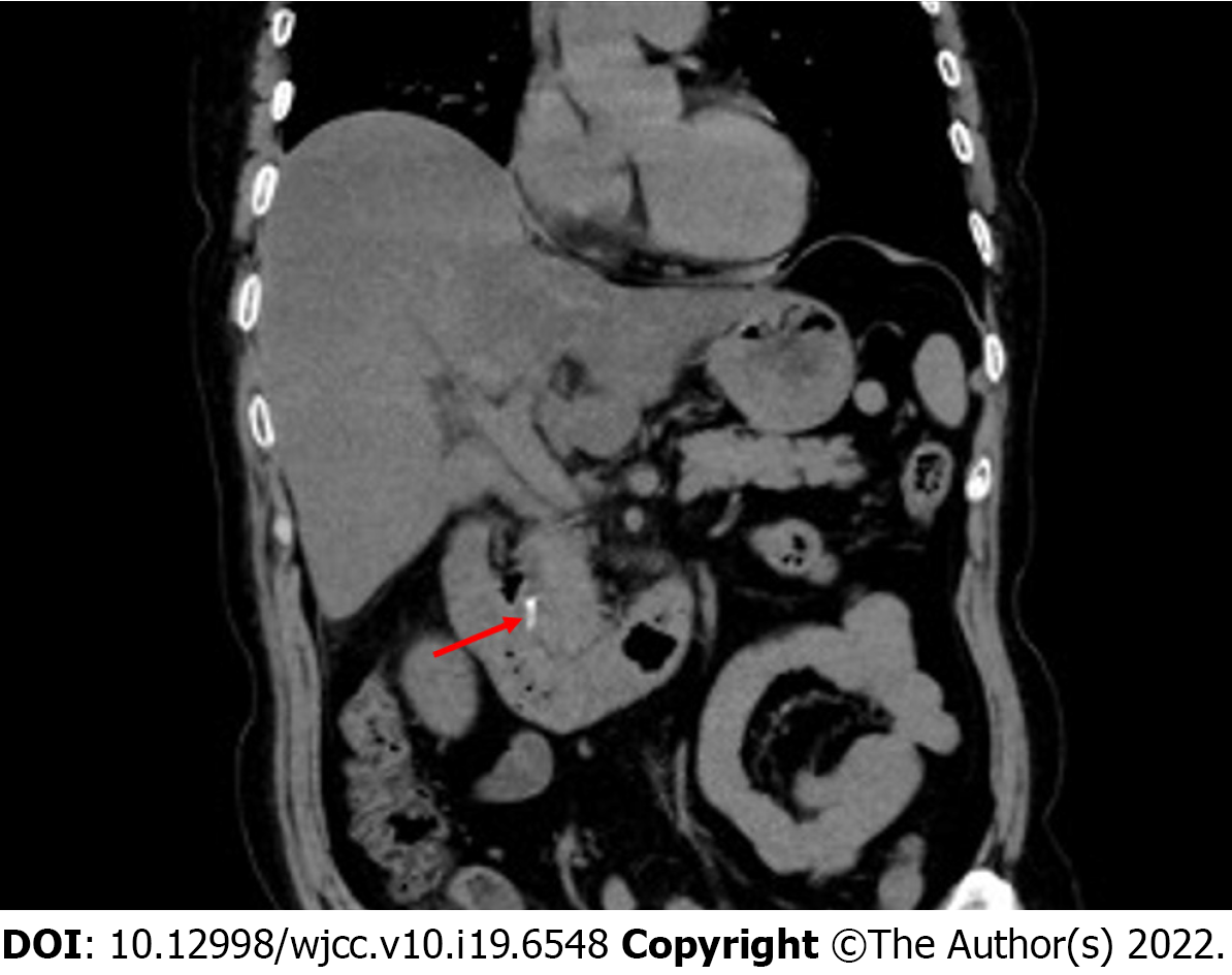
Figure 1 Abdominal computed tomography demonstrated hyperdense material in the common bile duct corresponding to the migrated Hem-o-lok clips.
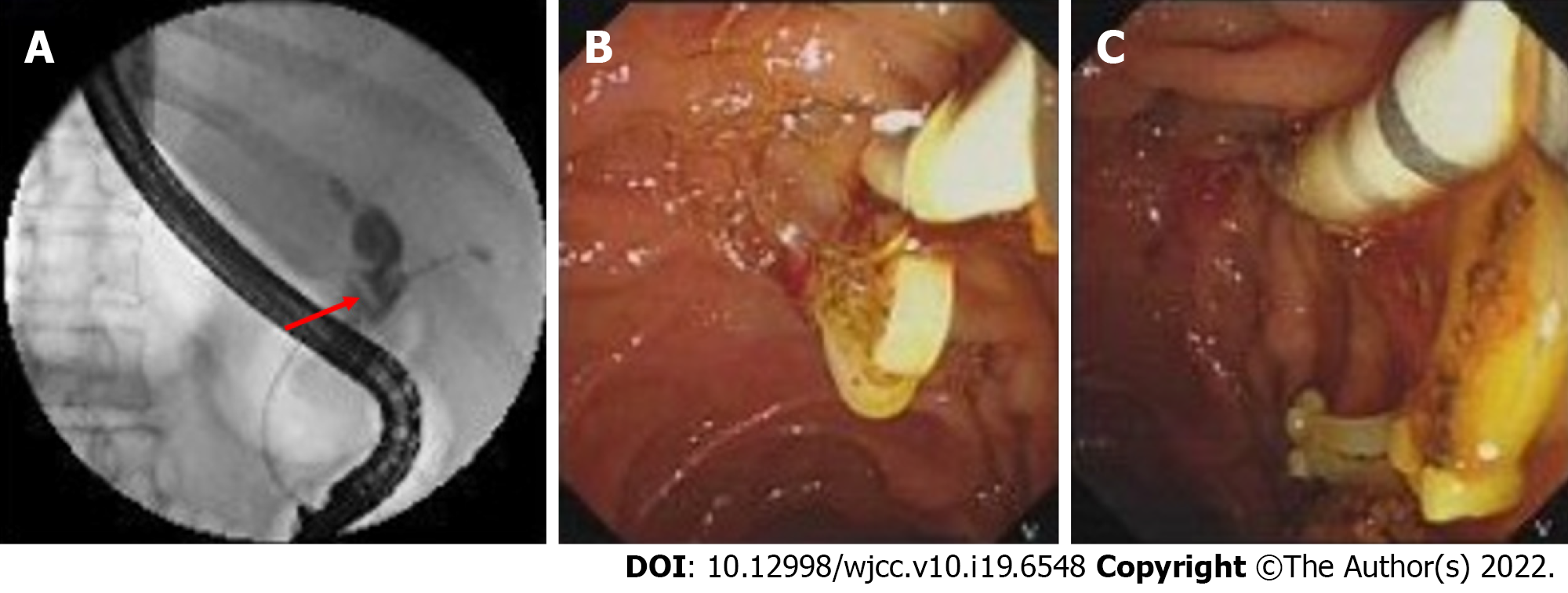
Figure 2 Migrated Hem-o-lok clips were detected and removed by endoscopic retrograde cholangiopancreatography.
A: Endoscopic retrograde cholangiopancreatography (ERCP) showed the filling-defect in the common bile duct corresponding to the migrated Hem-o-lok clips (red arrow); B and C: Hem-o-lok clips were removed by stone basket by ERCP.
- Citation: Liu DR, Wu JH, Shi JT, Zhu HB, Li C. Hem-o-lok clip migration to the common bile duct after laparoscopic common bile duct exploration: A case report. World J Clin Cases 2022; 10(19): 6548-6554
- URL: https://www.wjgnet.com/2307-8960/full/v10/i19/6548.htm
- DOI: https://dx.doi.org/10.12998/wjcc.v10.i19.6548


