Copyright
©The Author(s) 2022.
World J Clin Cases. Jul 6, 2022; 10(19): 6514-6519
Published online Jul 6, 2022. doi: 10.12998/wjcc.v10.i19.6514
Published online Jul 6, 2022. doi: 10.12998/wjcc.v10.i19.6514
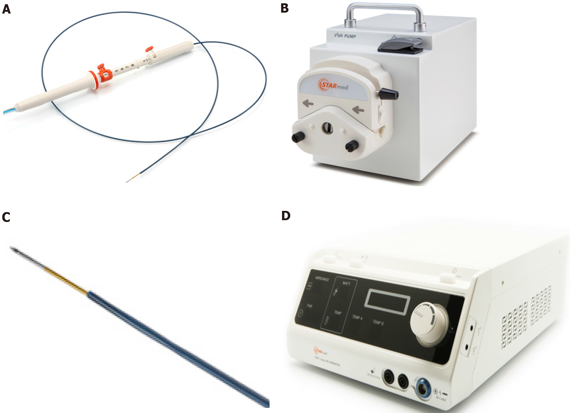
Figure 1 Radiofrequency ablation system.
A: Needle, similar to an endoscopic ultrasound fine needle aspiration or biopsy needle; B: Peristaltic pump which can infuse during the ablation, the electrode with chilled solution, maximizing volume ablation; C: Electrode on the distal needle tip, delivering radiofrequency ablation; D: Radiofrequency generator, with the possibility to monitor ablation parameters: Power, time, impedance. Citation: Rossi G, Petrone MC; Capurso G, Albarello L, Testoni SGG, Archibugi L, Lena MS, Doglioni C, Arcidiacono PG. Standardization of a Radiofrequency Ablation Tool in an Ex-Vivo Porcine Liver Model. Gastrointest Disord 2020; 2: 300-309. Copyright© The Authors 2020. Published by MDPI. No special permission is required to reuse all or part of article published by MDPI, including figures and tables, see https://www.mdpi.com/openaccess#Permissions. The authors have obtained the permission for figure using from Rossi G (Supplementary material).
Figure 2 Case 2 imaging.
A: A hyper-vascularized lesion compatible with an insulinoma, extremely close to the gastroduodenal artery is visible; B: Submucosal bleeding after radiofrequency ablation, treated by endoscopic hemostasis; C: Computed tomography scan 72 h after radiofrequency ablation: An 8 mm hypodense necrotic area at the previous lesion location, without signs of bleeding.
- Citation: Rossi G, Petrone MC, Capurso G, Partelli S, Falconi M, Arcidiacono PG. Endoscopic ultrasound radiofrequency ablation of pancreatic insulinoma in elderly patients: Three case reports. World J Clin Cases 2022; 10(19): 6514-6519
- URL: https://www.wjgnet.com/2307-8960/full/v10/i19/6514.htm
- DOI: https://dx.doi.org/10.12998/wjcc.v10.i19.6514


