Copyright
©The Author(s) 2022.
World J Clin Cases. May 16, 2022; 10(14): 4669-4675
Published online May 16, 2022. doi: 10.12998/wjcc.v10.i14.4669
Published online May 16, 2022. doi: 10.12998/wjcc.v10.i14.4669
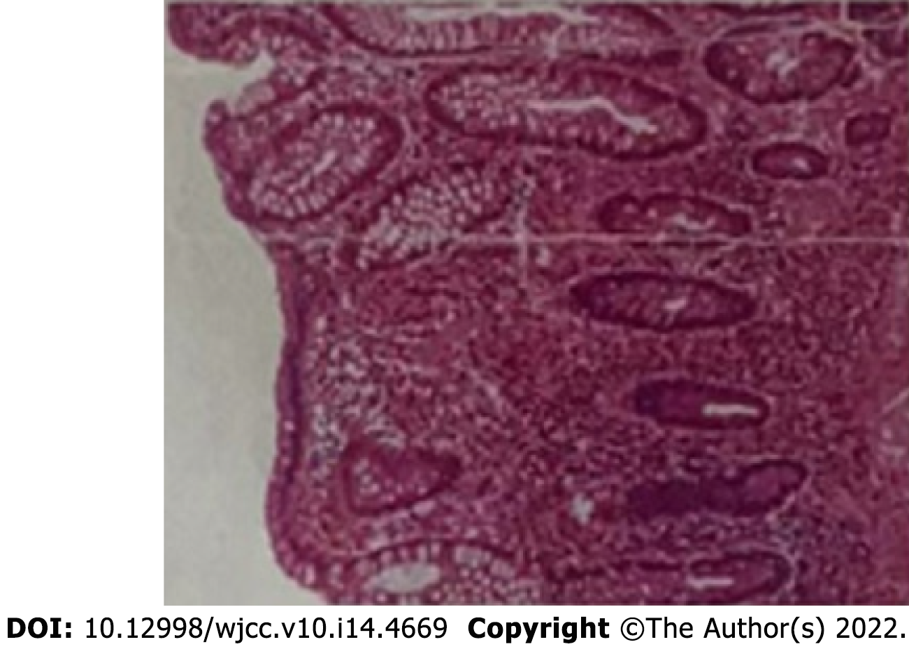
Figure 1 The pathology before neoadjuvant therapy.
Magnification × 100.
Figure 2 Computed tomography images of the patient (Colon hepatic flexure).
A: On the first abdominal computed tomography (CT) examination (2021-05), the intestinal wall at the hepatic curvature of the colon was significantly thickened with enhancement, the surrounding fat space was blurred, and the adjacent lymph nodes were enlarged; B: Abdominal CT examination (2021-07). The range of colonic lesions was reduced, and the surrounding lymph nodes were reduced; C: Abdominal CT examination (2021-09); the results showed improvement compared to the previous imaging results.
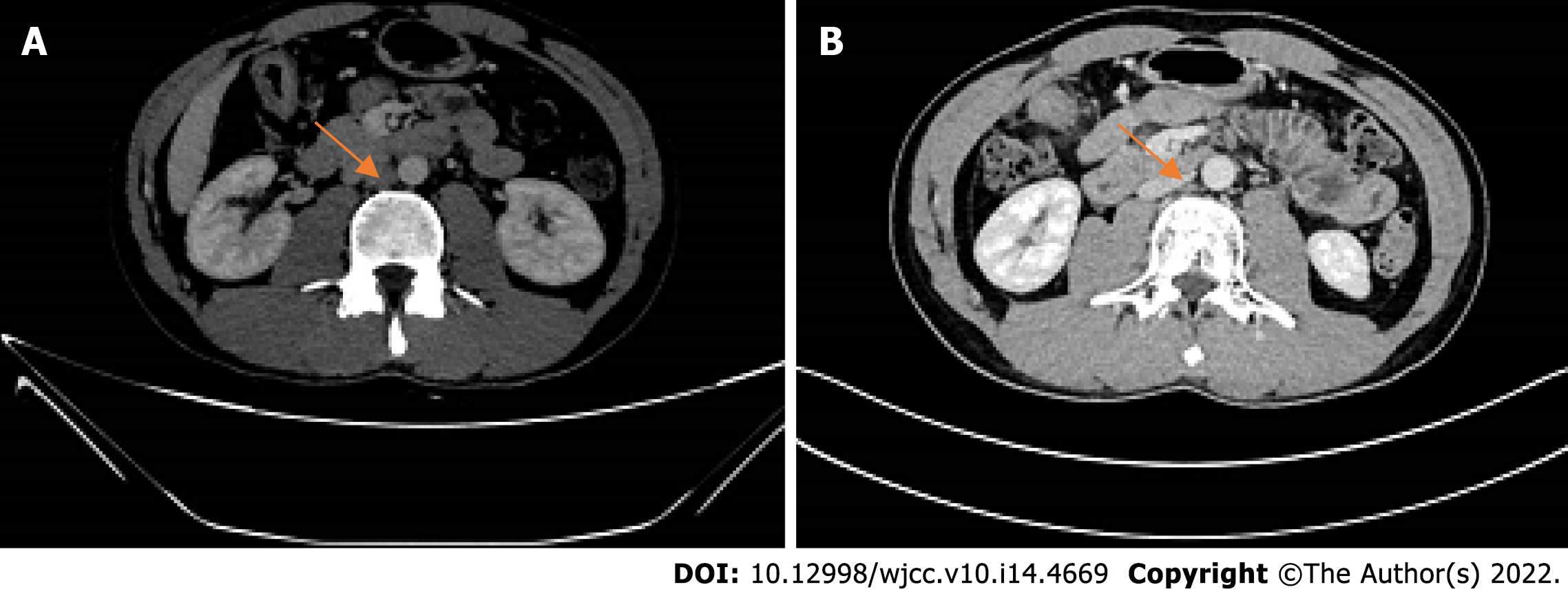
Figure 3 Computed tomography images of the patient (para-aortic lymph nodes).
A: Abdominal computed tomography (CT) examination (2021-05), enlarged lymph nodes beside the abdominal aorta; B: Abdominal CT examination (2021-07). The para-aortic lymph nodes were smaller than before.
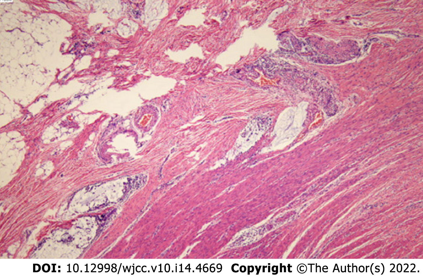
Figure 4 Haematoxylin-eosin staining and immunohistochemistry staining of the colon cancer tissue specimens.
Pathology after the neoadjuvant therapy. Magnification × 100.
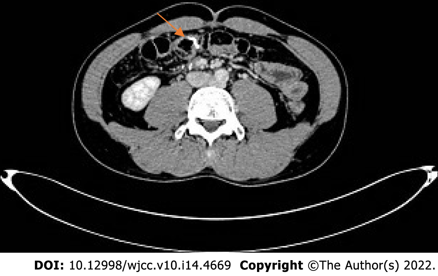
Figure 5
Abdominal computed tomography examination (2021-11): Postoperative review showed no evidence of recurrence.
- Citation: Zhang HQ, Huang CZ, Wu JY, Wang ZL, Shao Y, Fu Z. PD-1 inhibitor in combination with fruquintinib therapy for initial unresectable colorectal cancer: A case report. World J Clin Cases 2022; 10(14): 4669-4675
- URL: https://www.wjgnet.com/2307-8960/full/v10/i14/4669.htm
- DOI: https://dx.doi.org/10.12998/wjcc.v10.i14.4669


