Copyright
©The Author(s) 2022.
World J Clin Cases. May 16, 2022; 10(14): 4648-4653
Published online May 16, 2022. doi: 10.12998/wjcc.v10.i14.4648
Published online May 16, 2022. doi: 10.12998/wjcc.v10.i14.4648
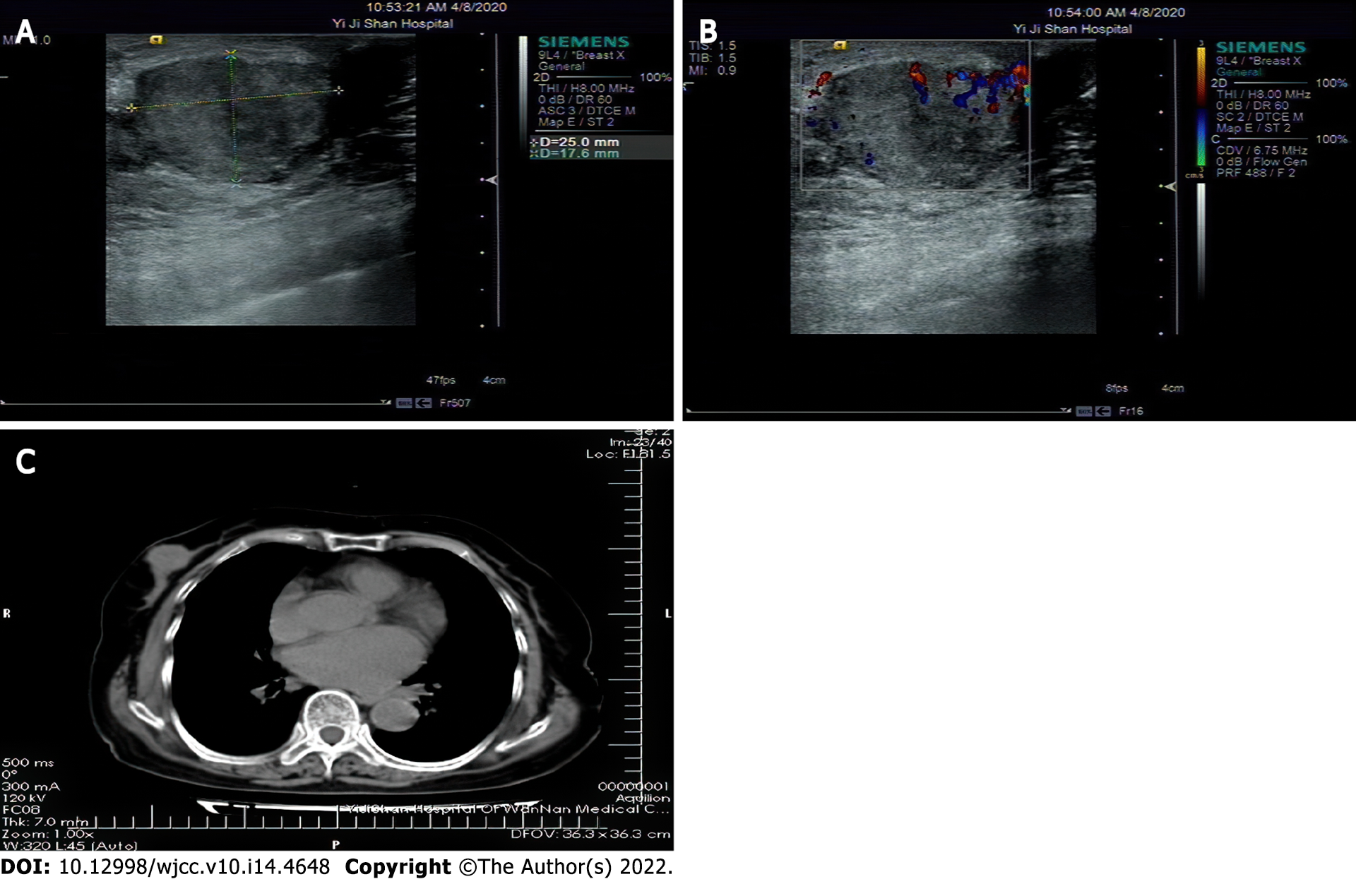
Figure 1 Breast ultrasound examination.
A: The lump and the surrounding boundary are clear, and the echo is uneven; B: Disturbance of blood flow signal in some areas, Breast Imaging Reporting and Data System score: 4C; C: Nodular soft tissue shadow in the right breast area.
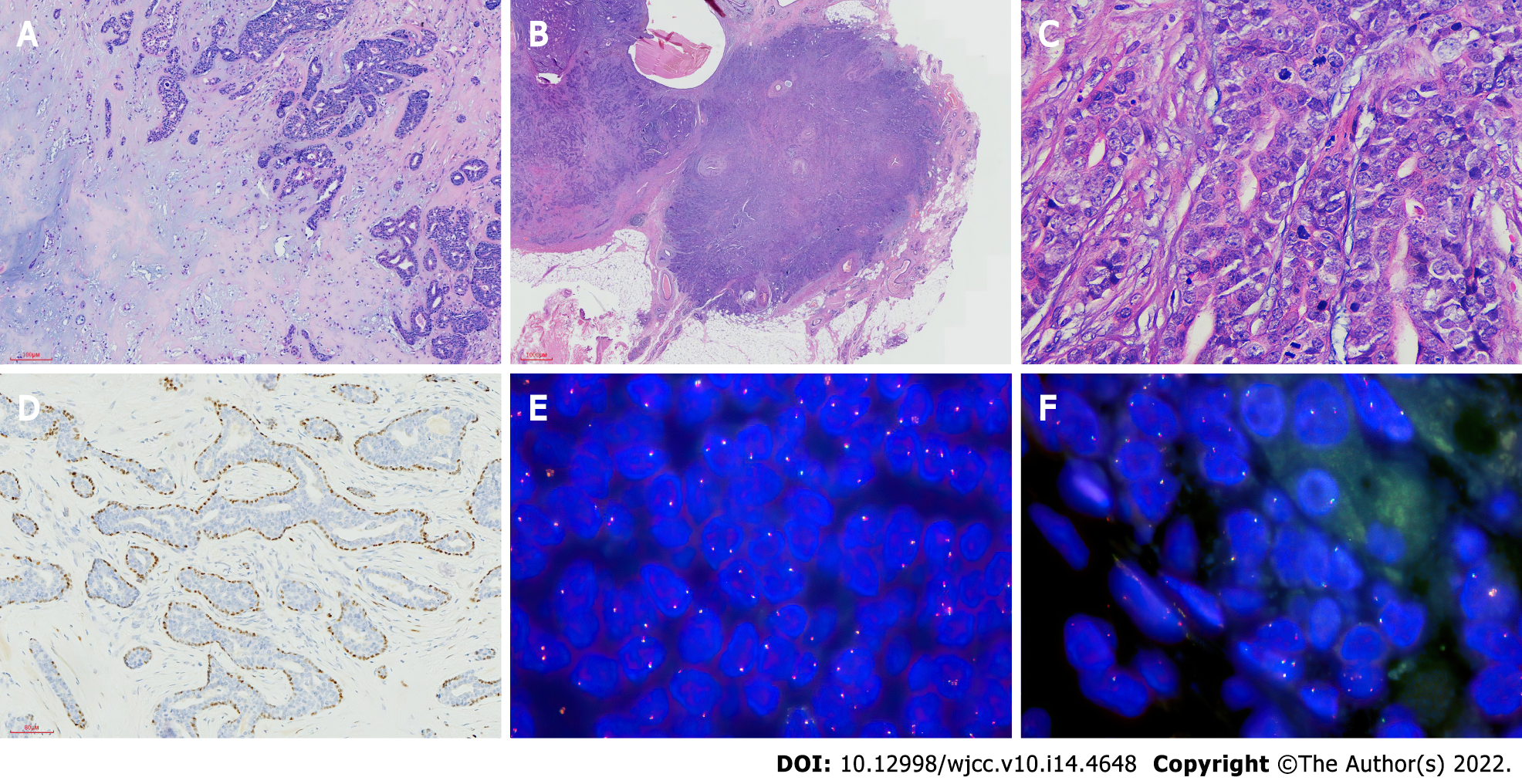
Figure 2 Pathological findings.
A: A glandular tubular structure is seen in the lesion, which contains pale eosin secretions × 200 HE; B: The tumor and the surrounding area are unclear in some areas, showing invasive growth × 100 HE; C: The tumor cells were heterotypic, with a high ratio of cytoplasm to nuclei; multiple mitotic figures × 400 HE; D: p63 shows intact myoepithelial × 200 IHC; E: HMGA2 fragmentation probe: Count 100 cells, 1R1G1F accounted for 1%, 1R1F accounted for 1%; F: PLAG1 fragmentation probe: Count 100 cells, 1R1G1F accounted for 2%.
- Citation: Zhang WT, Wang YB, Ang Y, Wang HZ, Li YX. Diagnosis of an extremely rare case of malignant adenomyoepithelioma in pleomorphic adenoma: A case report. World J Clin Cases 2022; 10(14): 4648-4653
- URL: https://www.wjgnet.com/2307-8960/full/v10/i14/4648.htm
- DOI: https://dx.doi.org/10.12998/wjcc.v10.i14.4648


