Copyright
©The Author(s) 2022.
World J Clin Cases. May 16, 2022; 10(14): 4494-4501
Published online May 16, 2022. doi: 10.12998/wjcc.v10.i14.4494
Published online May 16, 2022. doi: 10.12998/wjcc.v10.i14.4494
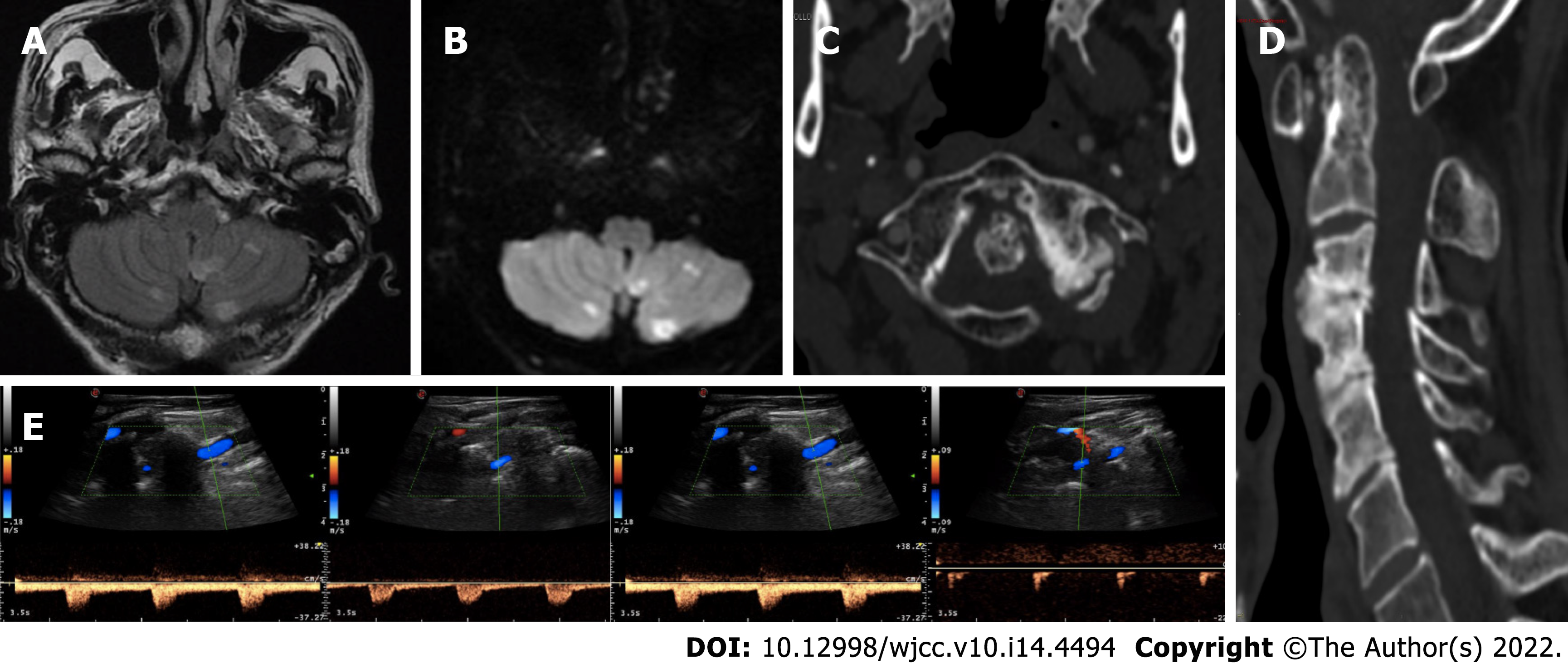
Figure 1 Neuroradiological and neurosonological evaluation.
A and B: Axial fluid-attenuated inversion-recovery and diffusion-weighted imaging (DWI) brain magnetic resonance imaging sequences with subacute bilateral cerebellar ischaemic lesions involving the area supplied by the posterior inferior cerebellar artery; C and D: Axial and sagittal cervical computed tomography scan showing marked degenerative joint alterations with atlo-axial instability and retroposition of dens, spinal canal stenosis and ankilosis of lateral left zygo-apophisal joints with underlying congenital partial atlo-occipital fusion; E: Vertebral ultrasound examination documenting regular blood flow in the left VA in the neutral position (left side) and “stump flow” demodulation in both the V1-V2 and V3-V4 segments (right side) in cases of slight contralateral head rotation (20°).
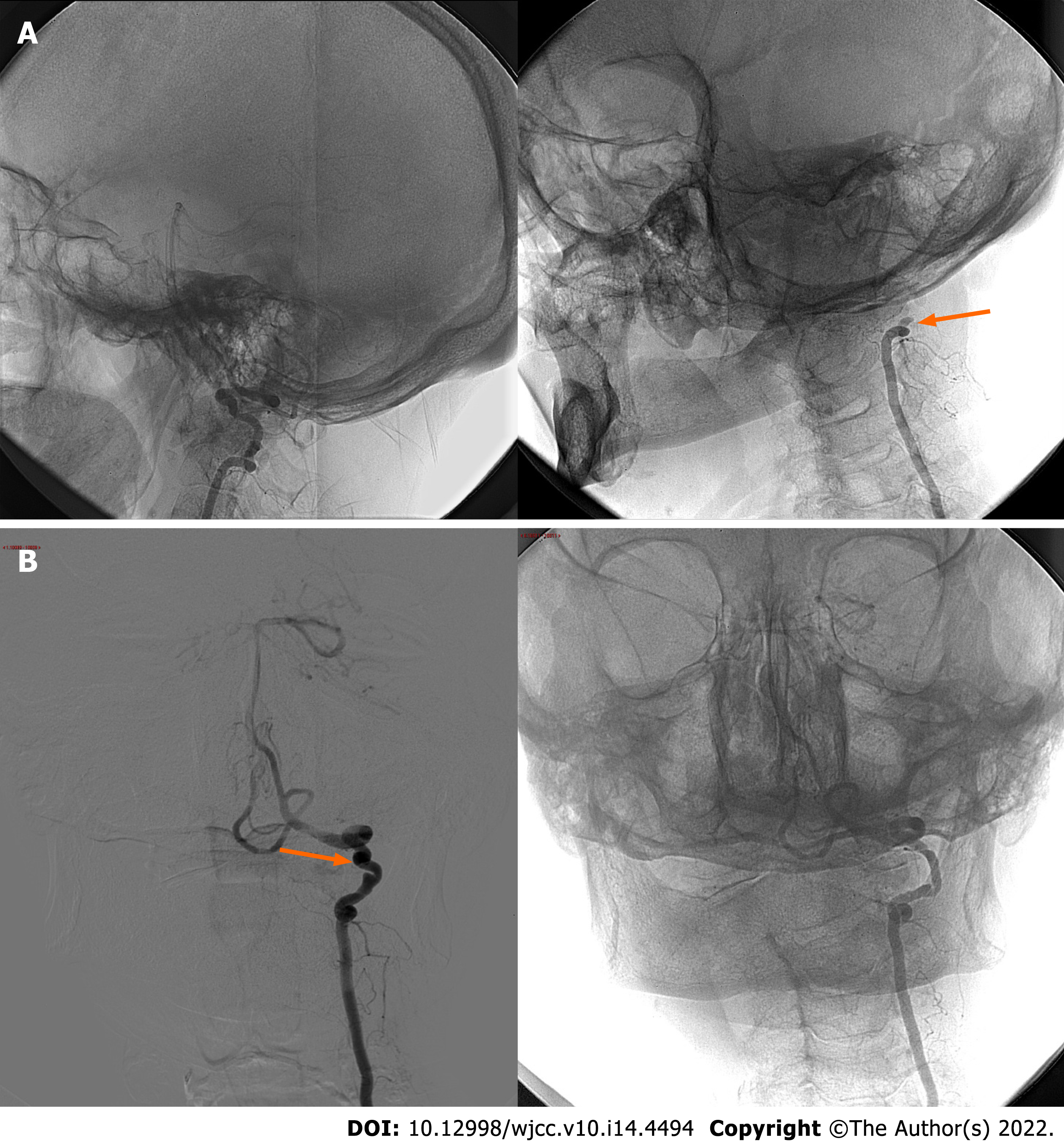
Figure 2 Digital subtraction angiography.
A: Lateral cerebral angiography projections without stenosis of the left vertebral artery in the neutral position (left side) and with complete occlusion of the V3 segment at the C2 Level upon turning the head to the right at 40° (right side; arrow); B: Anteroposterior cerebral angiography projections confirming the dynamic occlusion of the left VA in case of right head rotation (right side). Please note left VA irregular luminal injection and focal parietal ectasia in the neutral position (left side; arrow).
- Citation: Orlandi N, Cavallieri F, Grisendi I, Romano A, Ghadirpour R, Napoli M, Moratti C, Zanichelli M, Pascarella R, Valzania F, Zedde M. Bow hunter’s syndrome successfully treated with a posterior surgical decompression approach: A case report and review of literature. World J Clin Cases 2022; 10(14): 4494-4501
- URL: https://www.wjgnet.com/2307-8960/full/v10/i14/4494.htm
- DOI: https://dx.doi.org/10.12998/wjcc.v10.i14.4494


