Copyright
©The Author(s) 2022.
World J Clin Cases. May 16, 2022; 10(14): 4368-4379
Published online May 16, 2022. doi: 10.12998/wjcc.v10.i14.4368
Published online May 16, 2022. doi: 10.12998/wjcc.v10.i14.4368
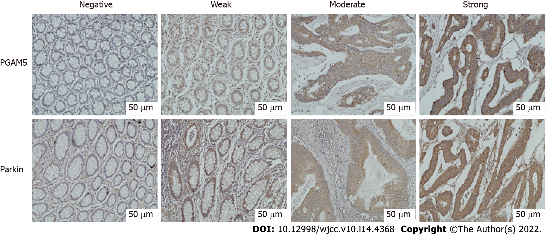
Figure 1 Representative immunohistochemical staining for phosphoglycerate mutase family member 5 and Parkin protein (original magnification 200 ×).
The expression levels of phosphoglycerate mutase family member 5 and Parkin protein were scored as negative (0), weak (1), moderate (2), or strong (3) according to staining intensity. PGAM5: Phosphoglycerate mutase family member 5.
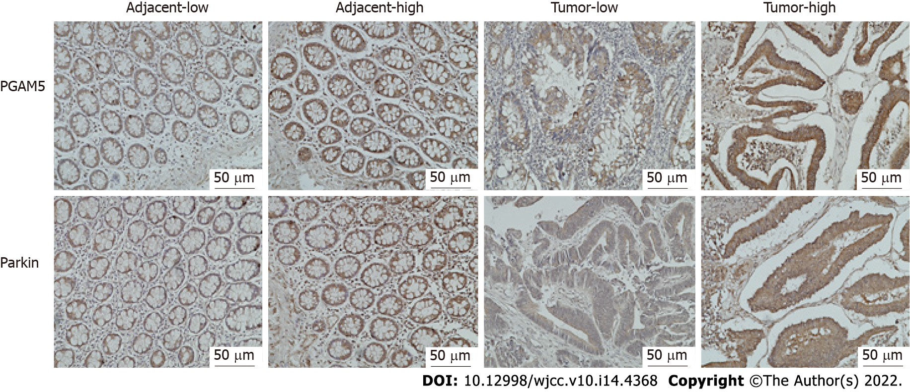
Figure 2 Representative immunohistochemical staining for phosphoglycerate mutase family member 5 and Parkin in colorectal adenocarcinoma tissues and adjacent tissues (original magnification 200 ×).
Phosphoglycerate mutase family member 5 (PGAM5) and Parkin protein were expressed in the cytoplasm of colonic epithelial cells. Specimens were categorized as either high or low expression according to the Youden index. High expression of PGAM5 was defined as a score of ≥ 4.75, and Parkin was ≥ 7.1. Low expression of PGAM5 was defined as a score of < 4.75, and Parkin was < 7.1. PGAM5: Phosphoglycerate mutase family member 5.
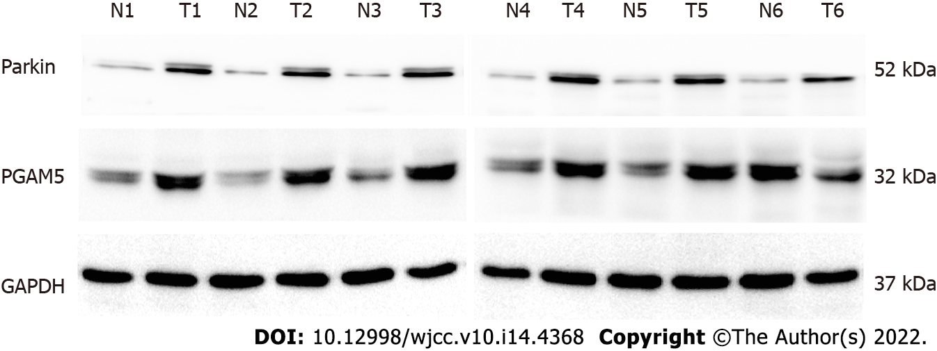
Figure 3 Western blots showing higher phosphoglycerate mutase family member 5 and Parkin levels in colorectal adenocarcinoma fresh tissues than in corresponding paracancerous tissues in six matched pairs (T, colorectal adenocarcinoma tissues; N, adjacent non-tumor tissues).
PGAM5: Phosphoglycerate mutase family member 5; GAPDH: Glyceraldehyde-3-phosphate dehydrogenase.
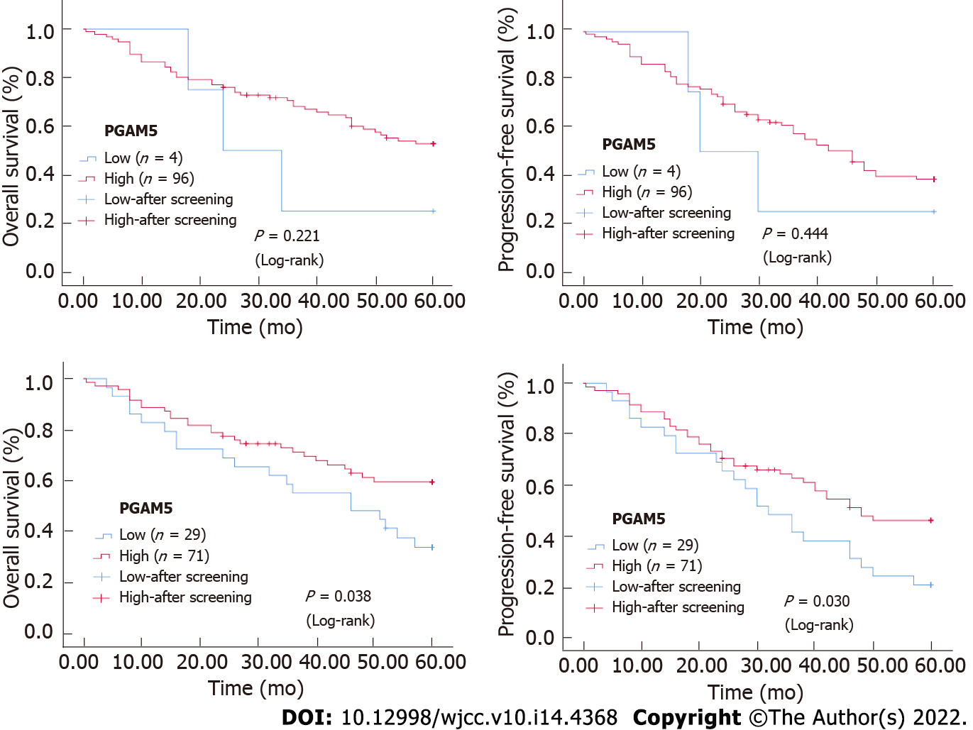
Figure 4 Kaplan-Meier survival curves for phosphoglycerate mutase family member 5 and Parkin expression in colorectal cancer tissues.
PGAM5: Phosphoglycerate mutase family member 5.
- Citation: Wu C, Feng ML, Jiao TW, Sun MJ. Clinical and prognostic significance of expression of phosphoglycerate mutase family member 5 and Parkin in advanced colorectal cancer. World J Clin Cases 2022; 10(14): 4368-4379
- URL: https://www.wjgnet.com/2307-8960/full/v10/i14/4368.htm
- DOI: https://dx.doi.org/10.12998/wjcc.v10.i14.4368


