Copyright
©The Author(s) 2018.
World J Methodol. Dec 14, 2018; 8(4): 51-58
Published online Dec 14, 2018. doi: 10.5662/wjm.v8.i4.51
Published online Dec 14, 2018. doi: 10.5662/wjm.v8.i4.51
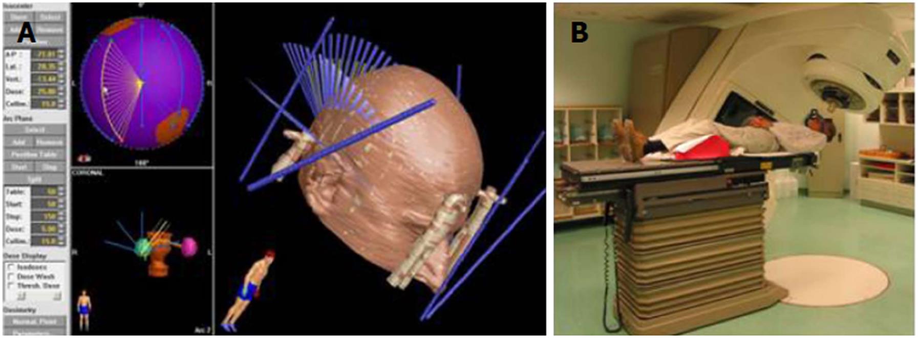
Figure 5 Irradiation with a linear accelerator.
A: A computer irradiation plan showing a localizer cube attached to the stereotactic frame (thick blue lines) (right image) and the direction of irradiation of the intracranial lesion with X-rays (thin blue lines). Small images on the left show the location of two intracranial lesions that are going to be irradiated; B: The patient is resting on the table with the head fixed in the stereotactic frame. The bed and gantry rotate about various axes, and this allows for irradiation by means of multiple trajectories intersecting at a certain point.
- Citation: Velnar T, Bosnjak R. Radiosurgical techniques for the treatment of brain neoplasms: A short review. World J Methodol 2018; 8(4): 51-58
- URL: https://www.wjgnet.com/2222-0682/full/v8/i4/51.htm
- DOI: https://dx.doi.org/10.5662/wjm.v8.i4.51


