Copyright
©The Author(s) 2018.
World J Methodol. Dec 14, 2018; 8(4): 51-58
Published online Dec 14, 2018. doi: 10.5662/wjm.v8.i4.51
Published online Dec 14, 2018. doi: 10.5662/wjm.v8.i4.51
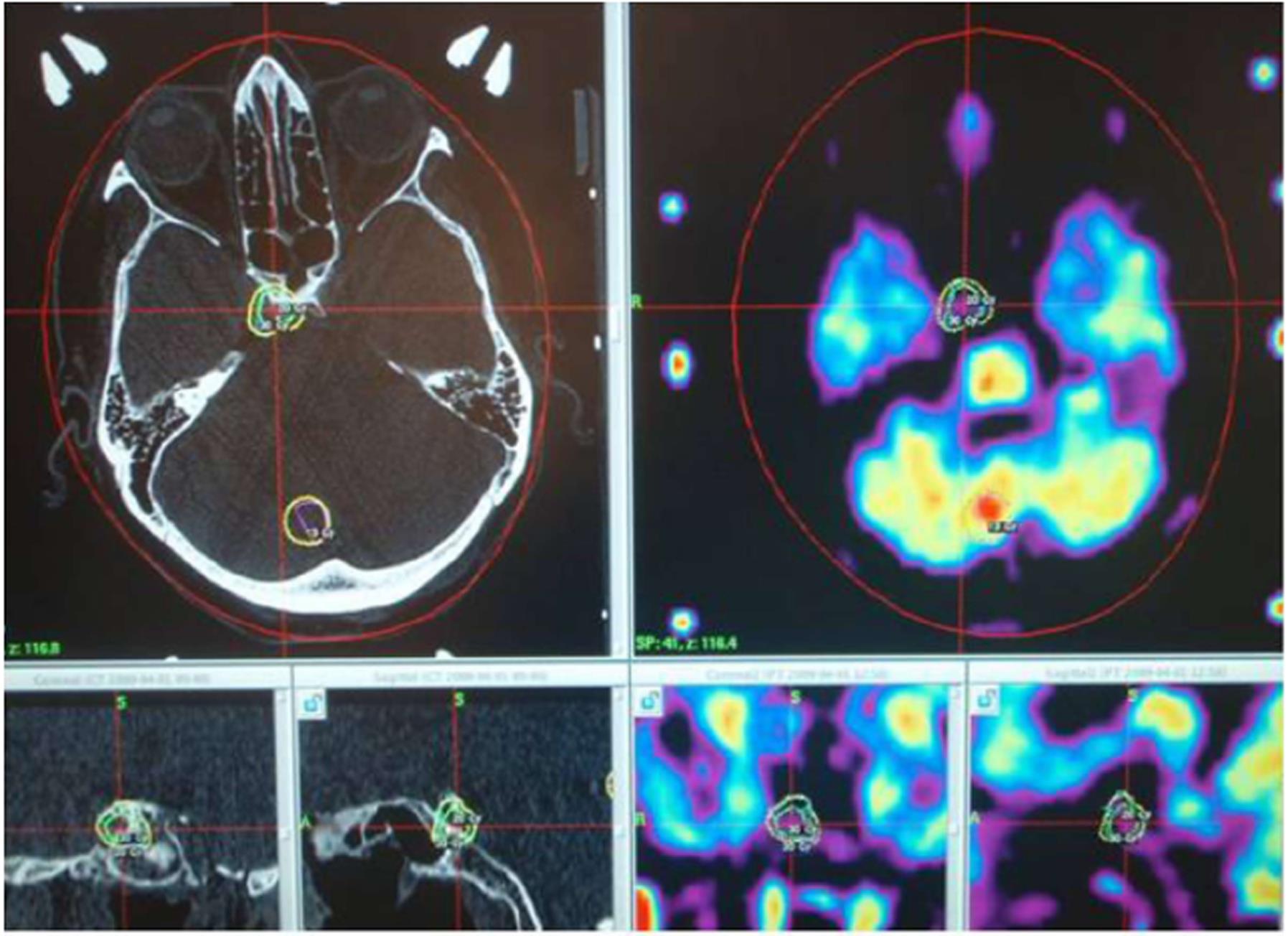
Figure 3 An example of computer planning for irradiation procedure of a residual pituitary adenoma, which was not completely removed from the parasellar space during a transnasal approach.
In the upper row are the axial views of CT and MRI-PET images with the tumor location. In the lower row from left to right: the frontal and sagittal views of CT and MRI-PET images. CT: Computed tomography; MRI-PET: Magnetic resonance imaging - positron emission tomography.
- Citation: Velnar T, Bosnjak R. Radiosurgical techniques for the treatment of brain neoplasms: A short review. World J Methodol 2018; 8(4): 51-58
- URL: https://www.wjgnet.com/2222-0682/full/v8/i4/51.htm
- DOI: https://dx.doi.org/10.5662/wjm.v8.i4.51


