Copyright
©The Author(s) 2017.
World J Methodol. Dec 26, 2017; 7(4): 117-128
Published online Dec 26, 2017. doi: 10.5662/wjm.v7.i4.117
Published online Dec 26, 2017. doi: 10.5662/wjm.v7.i4.117
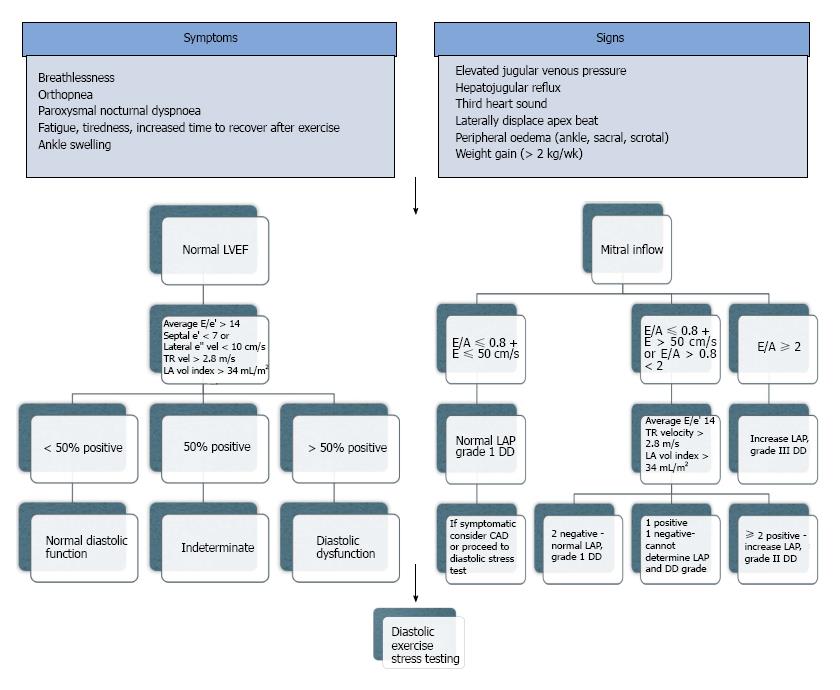
Figure 3 How to diagnose heart failure with preserved ejection fraction.
From the 2016 consensus statements of HF, the diagnosis of HF requires 4 important factors: (1) the presence of symptoms and/or signs of HF; (2) a “preserved” EF (defined as LVEF ≥ 50% or 40%-49% for HfmrEF; (3) elevated levels of natriuretic peptides (BNP > 35 pg/mL and/or NT-proBNP > 125 pg/mL); (4) objective evidence of other cardiac functional and structural alterations underlying HF; and (5) In case of uncertainty, a stress test or invasively measured elevated LV filling pressure may be needed to confirm the diagnosis. However in clinical practice many patients present predominately with a symptom such as SOB. The new guidelines are a positive step forward, and the authors for the first time acknowledged LA size, a surrogate for chronically elevated LVEDP and LA dysfunction. They fall short however as there are confounders for the abnormalities and none of the factors can be conclusively correlated to symptoms, where exercise testing could. A: Atrial filling velocity; BNP: Brain natriuretic peptides; E: Early filling velocity; e’: Early mitral annular tissue doppler velocity; EF: Ejection fraction; HfmrEF: Heart failure mid-range ejection fraction; LA: Left atrium; LAP: Left atrial pressure; LV: Left ventricle; LVEDP: Left ventricular end diastolic pressure; NT-proBNP: N Terminal Brain Natriuretic peptide; TR: Tricuspid regurgitation (adapted from References 1 and 3).
- Citation: Iyngkaran P, Anavekar NS, Neil C, Thomas L, Hare DL. Shortness of breath in clinical practice: A case for left atrial function and exercise stress testing for a comprehensive diastolic heart failure workup. World J Methodol 2017; 7(4): 117-128
- URL: https://www.wjgnet.com/2222-0682/full/v7/i4/117.htm
- DOI: https://dx.doi.org/10.5662/wjm.v7.i4.117


