Copyright
©The Author(s) 2017.
World J Methodol. Sep 26, 2017; 7(3): 108-111
Published online Sep 26, 2017. doi: 10.5662/wjm.v7.i3.108
Published online Sep 26, 2017. doi: 10.5662/wjm.v7.i3.108
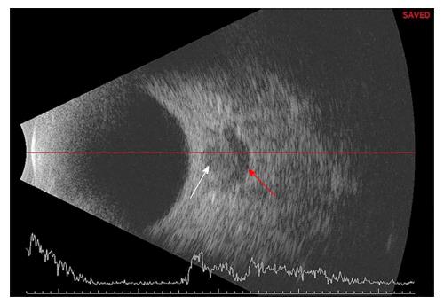
Figure 1 B scan ultrasound picture showing the normal round hypoechoic area of optic nerve (white arrow) with a hypoechoic crescent shaped area (red arrow) depicting “crescent” or “doughnut” sign.
- Citation: Bhosale A, Shah VM, Shah PK. Accuracy of crescent sign on ocular ultrasound in diagnosing papilledema. World J Methodol 2017; 7(3): 108-111
- URL: https://www.wjgnet.com/2222-0682/full/v7/i3/108.htm
- DOI: https://dx.doi.org/10.5662/wjm.v7.i3.108


