Copyright
©The Author(s) 2016.
World J Methodol. Mar 26, 2016; 6(1): 87-92
Published online Mar 26, 2016. doi: 10.5662/wjm.v6.i1.87
Published online Mar 26, 2016. doi: 10.5662/wjm.v6.i1.87
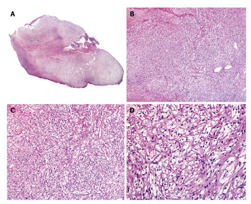
Figure 2 Histopathology of cutaneous perivascular epithelioid cell tumor.
A: Low power image of cutaneous perivascular epithelioid cell tumor; B and C: Medium power image showing the lobular arrangement of a neoplasm composed by clear cells; D: Detail of the cells with an ovoid homogeneous nuclei and a clear cytoplasm.
- Citation: Llamas-Velasco M, Requena L, Mentzel T. Cutaneous perivascular epithelioid cell tumors: A review on an infrequent neoplasm. World J Methodol 2016; 6(1): 87-92
- URL: https://www.wjgnet.com/2222-0682/full/v6/i1/87.htm
- DOI: https://dx.doi.org/10.5662/wjm.v6.i1.87


