Copyright
©2013 Baishideng Publishing Group Co.
World J Methodol. Dec 26, 2013; 3(4): 45-64
Published online Dec 26, 2013. doi: 10.5662/wjm.v3.i4.45
Published online Dec 26, 2013. doi: 10.5662/wjm.v3.i4.45
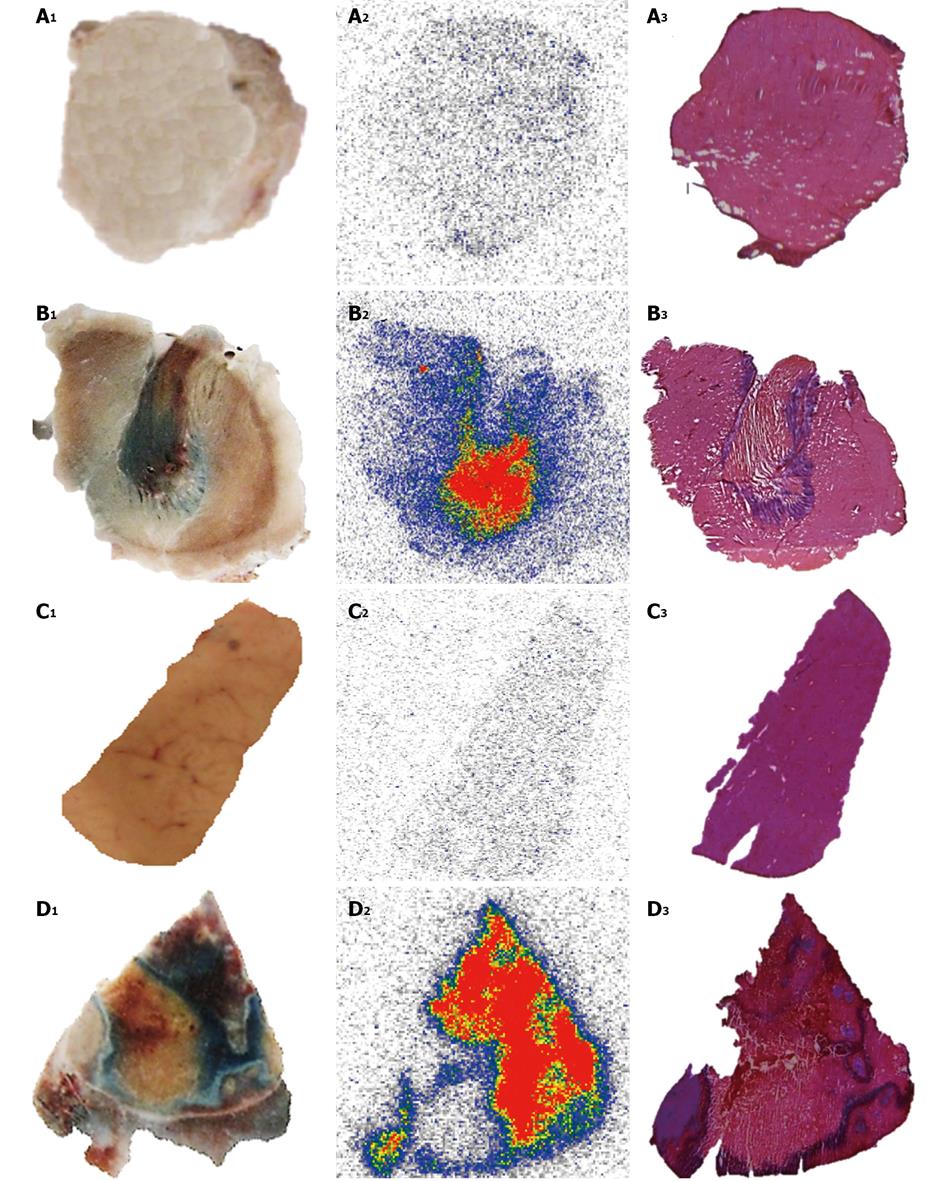
Figure 7 Post-mortem study of necrotic and viable tissues in the liver and muscle from animal models either of reperfused partial liver infarction or ethanol-induced muscle necrosis pre-injected with iodine-123 labeled hypericin/hypericin followed by 1% Evans blue solution.
A, B: Muscle; C, D: Liver. The hepatic infarction (A1) and necrotic muscle (B1) retain Evans blue as blue hyper intense areas, with viable liver (C1) and normal muscle (D1) without staining. Autoradiograms of 50-μm-thick sections show high tracer uptake in hepatic infarction (A2) and muscle necrosis (B2) but low accumulation either in viable liver (C2) or muscle (D2). The color code bar represents the code for the radioactivity concentration. By histology, the presence of hepatic infarction (A3) and muscle necrosis (B3) and the location of the viable liver (C3) and muscle (D3) tissues are verified.
- Citation: Cona MM, Witte P, Verbruggen A, Ni Y. An overview of translational (radio)pharmaceutical research related to certain oncological and non-oncological applications. World J Methodol 2013; 3(4): 45-64
- URL: https://www.wjgnet.com/2222-0682/full/v3/i4/45.htm
- DOI: https://dx.doi.org/10.5662/wjm.v3.i4.45


