Copyright
©2013 Baishideng Publishing Group Co.
World J Methodol. Sep 26, 2013; 3(3): 27-38
Published online Sep 26, 2013. doi: 10.5662/wjm.v3.i3.27
Published online Sep 26, 2013. doi: 10.5662/wjm.v3.i3.27
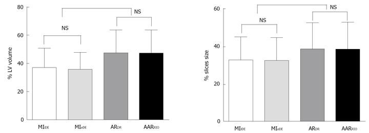
Figure 7 Comparisons between myocardial infarction and area at risk by different techniques.
There were no significant differences between myocardial infarction in vivo delayed enhancement (MI iDE) and myocardial infarction ex vivo delayed enhancement (MI eDE) measured from cardiac magnetic resonance imaging in global (left) and slices (right). There were no significant differences between area at risk digital radiography (AARDR) and area at risk red iodized oil-staining (AARRIO-staining) in global (left) and slices (right).The significant differences were observed on AARDRvs MI iDE (P < 0.01); AARDRvs MI eDE (P < 0.01); AARRIO-stainingvs MI iDE (P < 0.01); AARRIO-stainingvs MI eDE (P < 0.01). NS: Not significant.
-
Citation: Feng Y, Ma ZL, Chen F, Yu J, Cona MM, Xie Y, Li Y, Ni Y. Bifunctional staining for
ex vivo determination of area at risk in rabbits with reperfused myocardial infarction. World J Methodol 2013; 3(3): 27-38 - URL: https://www.wjgnet.com/2222-0682/full/v3/i3/27.htm
- DOI: https://dx.doi.org/10.5662/wjm.v3.i3.27


