Copyright
©2013 Baishideng Publishing Group Co.
World J Methodol. Sep 26, 2013; 3(3): 27-38
Published online Sep 26, 2013. doi: 10.5662/wjm.v3.i3.27
Published online Sep 26, 2013. doi: 10.5662/wjm.v3.i3.27
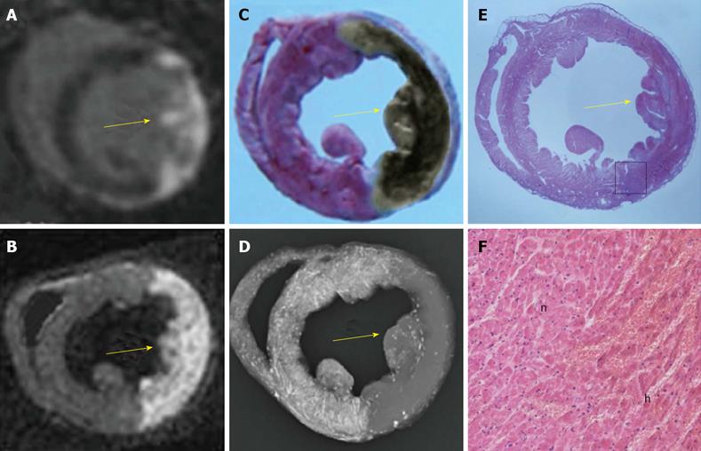
Figure 4 An example of a reperfused hemorrhagic myocardial infarction (arrow) shown by cardiac magnetic resonance imaging and the area at risk demonstrated with bifunctional staining method.
A: The hyperenhanced transmural myocardial infarction (MI) was seen at the lateral wall on in vivo delay enhancement (DE) cardiac magnetic resonance imaging (cMRI); B: The ex vivo DE cMRI was highly correspondent with A; C: In contrast to the normal myocardium with red staining, the area at risk (AAR) was shown as an unstained region, on which the brown grayish area indicates a large intramural hemorrhagic infarction after the fixation by Formalin; D: On digital radiography (DR), the AAR was shown as non-opacified region in contrast to the opacified normal myocardium. Notice that the AAR defined by red iodized oil-staining (RIO-staining) in C and DR in D was somewhat larger than the MI defined by in vivo DE cMRI in A and ex vivo DE cMRI in B; E: The hematoxylin-eosin (HE) stained macroscopic view was obtained from the corresponding histological section of A, B, C, and D; F: Photomicroscopic view of the HE stained slice (magnification, × 100) confirmed the presence of myocardial necrosis with hemorrhage and tissue reaction. h: Hemorrhagic infarction; n: Adjacent normal myocardium with inflammatory infiltration.
-
Citation: Feng Y, Ma ZL, Chen F, Yu J, Cona MM, Xie Y, Li Y, Ni Y. Bifunctional staining for
ex vivo determination of area at risk in rabbits with reperfused myocardial infarction. World J Methodol 2013; 3(3): 27-38 - URL: https://www.wjgnet.com/2222-0682/full/v3/i3/27.htm
- DOI: https://dx.doi.org/10.5662/wjm.v3.i3.27


