Copyright
©2013 Baishideng Publishing Group Co.
World J Methodol. Sep 26, 2013; 3(3): 27-38
Published online Sep 26, 2013. doi: 10.5662/wjm.v3.i3.27
Published online Sep 26, 2013. doi: 10.5662/wjm.v3.i3.27
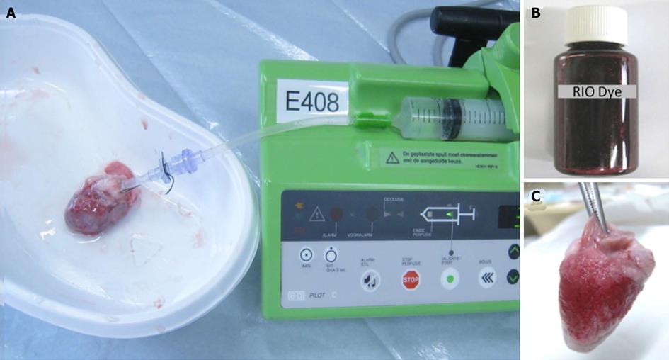
Figure 3 Bifunctional staining methods.
A: A plastic catheter filled with saline was inserted into the aorta and anchored with its tip 1 cm above the aortic valves, which was connected to a 10 mL syringe fixed in the injection bump. The heart was gently rinsed to wash out remaining blood, then the left circumflex coronary artery was re-ligated, and 2-4 mL of red iodized oil (RIO) dye was infused; B: Homemade RIO dye; C: Lateral view of the RIO perfused heart. The perfusion of RIO dye was stopped when the normal myocardium was stained completely scarlet red, while the area at risk remained unstained.
-
Citation: Feng Y, Ma ZL, Chen F, Yu J, Cona MM, Xie Y, Li Y, Ni Y. Bifunctional staining for
ex vivo determination of area at risk in rabbits with reperfused myocardial infarction. World J Methodol 2013; 3(3): 27-38 - URL: https://www.wjgnet.com/2222-0682/full/v3/i3/27.htm
- DOI: https://dx.doi.org/10.5662/wjm.v3.i3.27


