Copyright
©The Author(s) 2024.
World J Methodol. Dec 20, 2024; 14(4): 95685
Published online Dec 20, 2024. doi: 10.5662/wjm.v14.i4.95685
Published online Dec 20, 2024. doi: 10.5662/wjm.v14.i4.95685
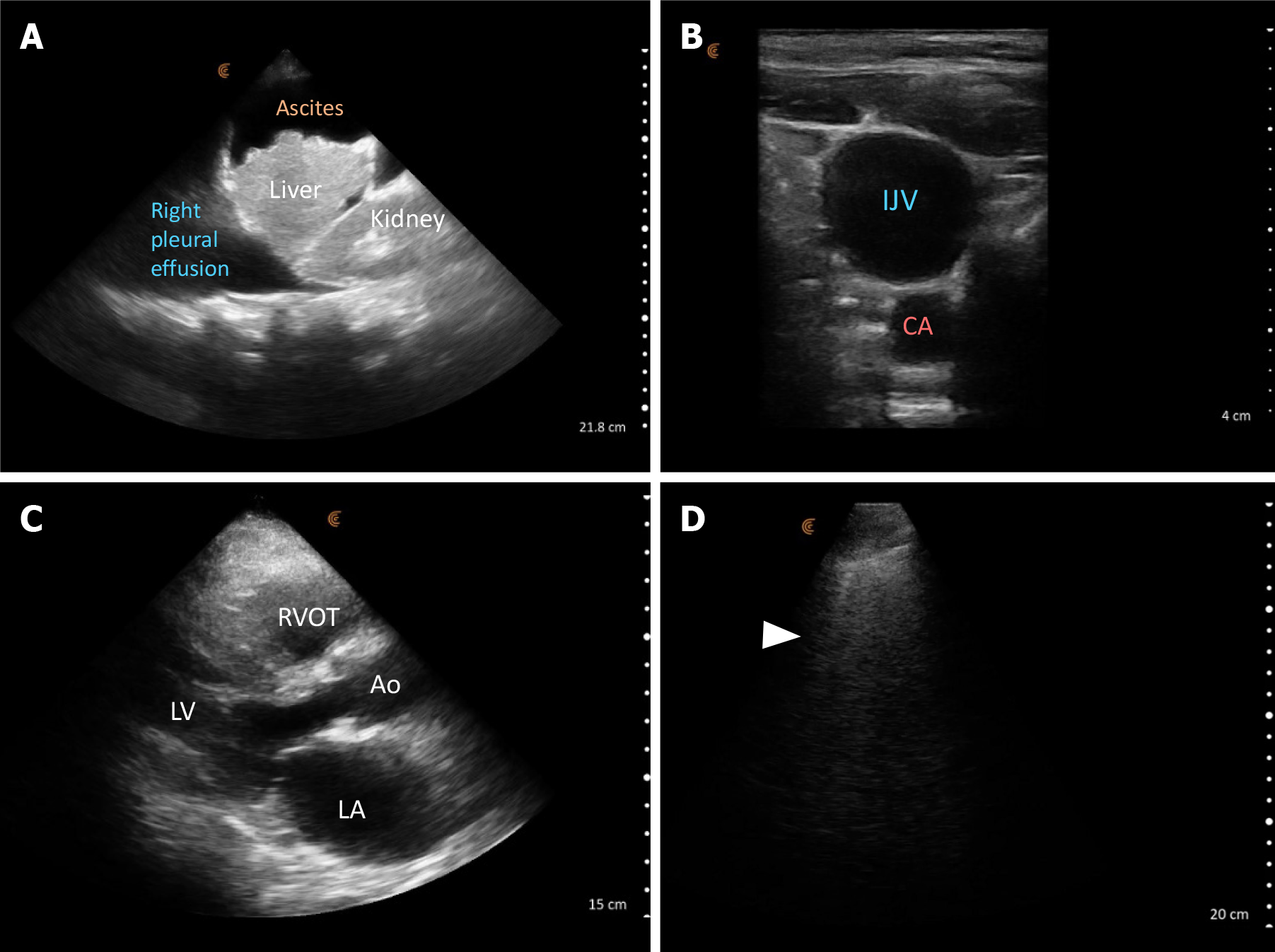
Figure 7 Point-of-care ultrasound images demonstrating findings suggestive of elevated cardiac filling pressures.
A: Right pleural effusion and ascites as seen on right upper quadrant lateral scan plane; B: A dilated internal jugular vein (IJV) at the cricoid cartilage level with a head angle of approximately 45 degrees consistent with elevated right atrial pressure. Adjacent carotid artery (CA) is seen: C: Parasternal long axis cardiac view showing a qualitatively dilated left atrium; D: Lung ultrasound image demonstrating vertical B-lines (arrowhead) indicative of elevated extravascular lung water. LA: left atrium; Ao: Aorta; LV: Left ventricle; RVOT: Right ventricular outflow tract.
- Citation: Sinanan R, Moshtaghi A, Koratala A. Point-of-care ultrasound in nephrology: A private practice viewpoint. World J Methodol 2024; 14(4): 95685
- URL: https://www.wjgnet.com/2222-0682/full/v14/i4/95685.htm
- DOI: https://dx.doi.org/10.5662/wjm.v14.i4.95685


