Copyright
©The Author(s) 2024.
World J Methodol. Dec 20, 2024; 14(4): 95685
Published online Dec 20, 2024. doi: 10.5662/wjm.v14.i4.95685
Published online Dec 20, 2024. doi: 10.5662/wjm.v14.i4.95685
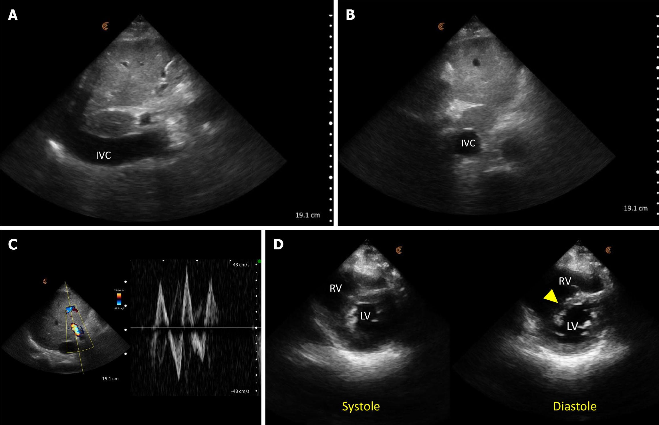
Figure 5 Point-of-care ultrasound images demonstrating findings suggestive of volume/pressure overload.
A: A dilated inferior vena cava (IVC) in long axis; B: IVC transverse view shows a circular vessel as opposed to normal elliptical shape; C: Pulsatile portal vein Doppler with a to-and-fro pattern (normally it’s continuous); D: Parasternal short axis cardiac view showing interventricular septal flattening (arrowhead) predominantly in diastole. RV: Right ventricle; LV: Left ventricle.
- Citation: Sinanan R, Moshtaghi A, Koratala A. Point-of-care ultrasound in nephrology: A private practice viewpoint. World J Methodol 2024; 14(4): 95685
- URL: https://www.wjgnet.com/2222-0682/full/v14/i4/95685.htm
- DOI: https://dx.doi.org/10.5662/wjm.v14.i4.95685


