Copyright
©The Author(s) 2024.
World J Methodol. Dec 20, 2024; 14(4): 95685
Published online Dec 20, 2024. doi: 10.5662/wjm.v14.i4.95685
Published online Dec 20, 2024. doi: 10.5662/wjm.v14.i4.95685
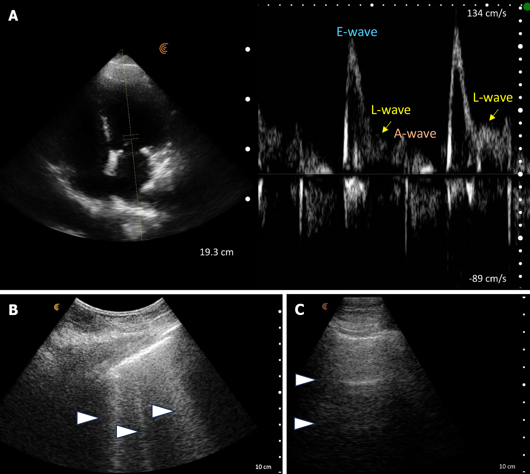
Figure 3 Point-of-care ultrasound images at the time of nephrology consult.
A: An elevated E-wave to A-wave ratio and an L-wave on transmitral Doppler; B: Vertical B-lines on lung ultrasound at initial examination (arrowheads). Follow up lung ultrasound; C: Horizontal A-lines (arrowheads) on lung ultrasound - normal finding.
- Citation: Sinanan R, Moshtaghi A, Koratala A. Point-of-care ultrasound in nephrology: A private practice viewpoint. World J Methodol 2024; 14(4): 95685
- URL: https://www.wjgnet.com/2222-0682/full/v14/i4/95685.htm
- DOI: https://dx.doi.org/10.5662/wjm.v14.i4.95685


