Copyright
©The Author(s) 2023.
World J Methodol. Sep 20, 2023; 13(4): 366-372
Published online Sep 20, 2023. doi: 10.5662/wjm.v13.i4.366
Published online Sep 20, 2023. doi: 10.5662/wjm.v13.i4.366
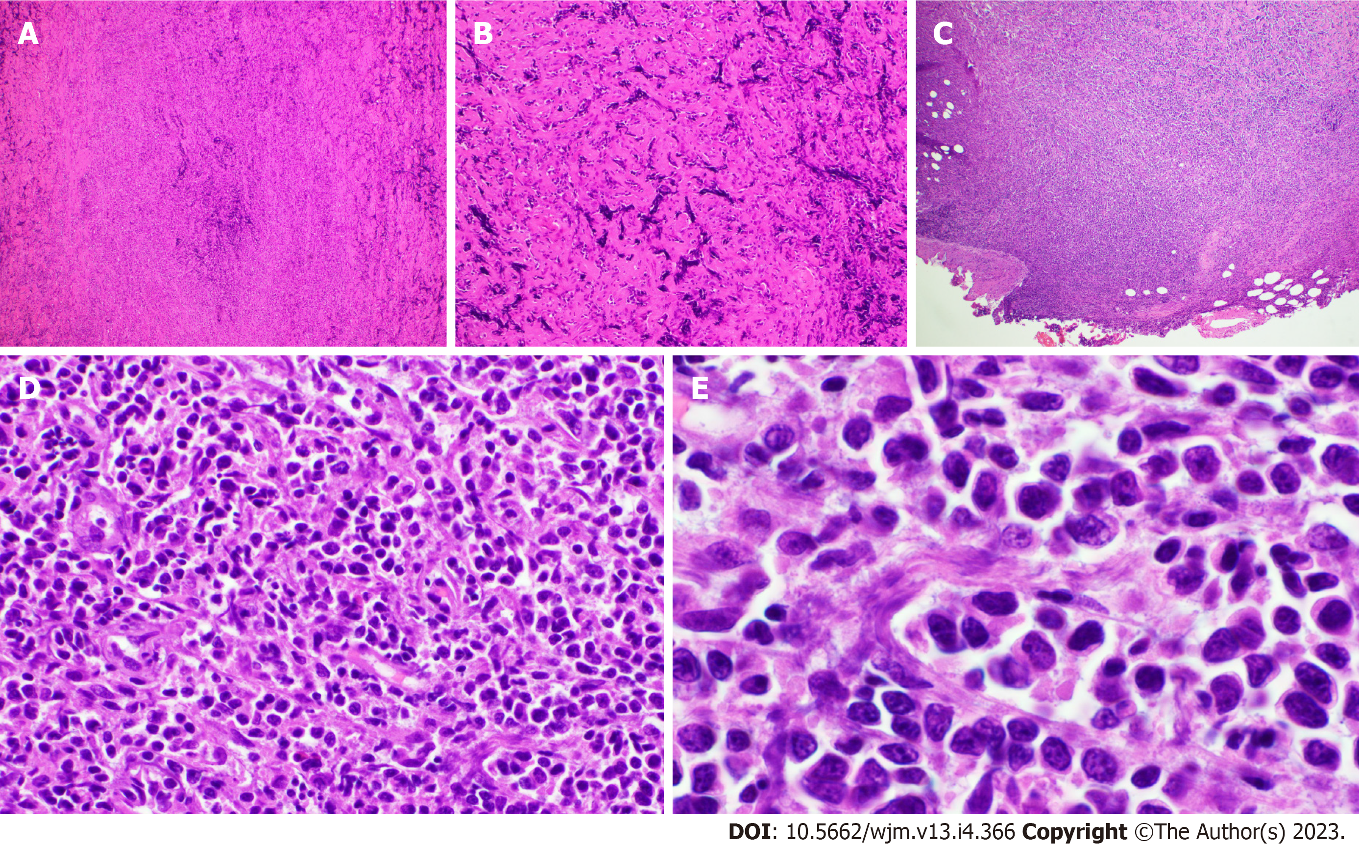
Figure 1 Histology of sclerotic marginal zone lymphoma.
A: Low power hematoxylin and eosin (H&E) section showing extensive fibrosis and crush artifact, 40 × (Olympus BX43); B: Low power H&E section showing extensive fibrosis and crush artifact, 40 × (Olympus BX43); C: More cellular area with small lymphocytes is seen focally (lower-left hand) compared with more sclerotic pattern (upper-right), 40 ×; D: High power H&E showing morphology of cells in cellular area (non-crushed), some have monocytoid features (400 ×); E: High power H&E showing morphology of cells in cellular area (non-crushed), some have monocytoid features (1000 ×).
- Citation: Moureiden Z, Tashkandi H, Hussaini MO. Sclerotic marginal zone lymphoma: A case report. World J Methodol 2023; 13(4): 366-372
- URL: https://www.wjgnet.com/2222-0682/full/v13/i4/366.htm
- DOI: https://dx.doi.org/10.5662/wjm.v13.i4.366


