Copyright
©The Author(s) 2023.
World J Methodol. Sep 20, 2023; 13(4): 359-365
Published online Sep 20, 2023. doi: 10.5662/wjm.v13.i4.359
Published online Sep 20, 2023. doi: 10.5662/wjm.v13.i4.359
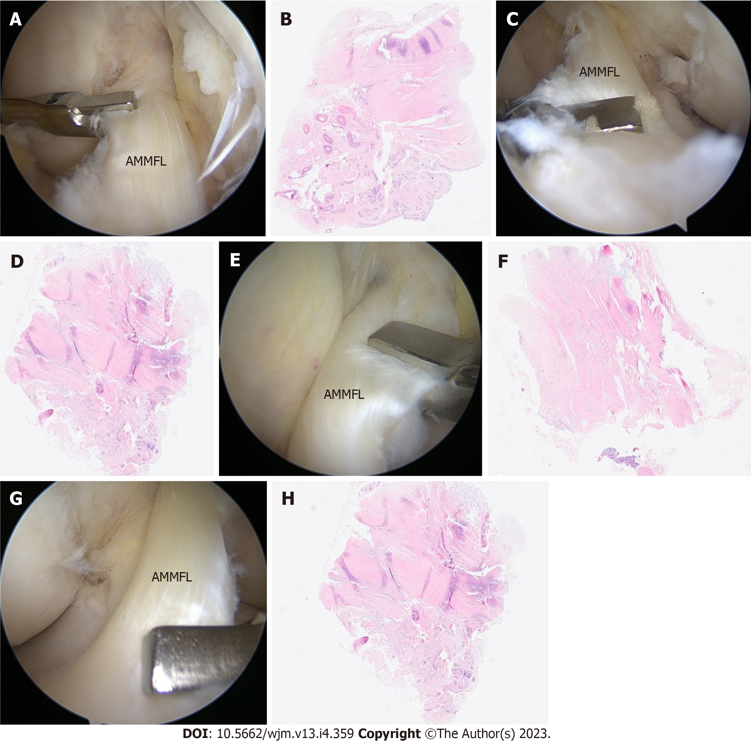
Figure 3 Intraoperative images and histologic examination of the knee.
A-D: Intraoperative images and histologic examination of the right knee. Arthroscopic images obtained through the anteromedial portal showing the biopsies performed through the anterolateral portal (A and B); hematoxylin and eosin staining of the meniscofemoral band reveals fibrocartilaginous tissue compatible with meniscus in both cases (C and D); E-H: Intraoperative images and histologic examination of the left knee. Arthroscopic images obtained through the anteromedial portal showing the biopsies performed through the anterolateral portal (E and F); hematoxylin and eosin staining of the meniscofemoral band reveals fibrocartilaginous tissue compatible with meniscus in both cases (G and H). AMMFL: Anteromedial meniscofemoral ligament.
- Citation: Luco JB, Di Memmo D, Gomez Sicre V, Nicolino TI, Costa-Paz M, Astoul J, Garcia-Mansilla I. Clinical, imaging, arthroscopic, and histologic features of bilateral anteromedial meniscofemoral ligament: A case report. World J Methodol 2023; 13(4): 359-365
- URL: https://www.wjgnet.com/2222-0682/full/v13/i4/359.htm
- DOI: https://dx.doi.org/10.5662/wjm.v13.i4.359


