Copyright
©The Author(s) 2023.
World J Methodol. Sep 20, 2023; 13(4): 359-365
Published online Sep 20, 2023. doi: 10.5662/wjm.v13.i4.359
Published online Sep 20, 2023. doi: 10.5662/wjm.v13.i4.359
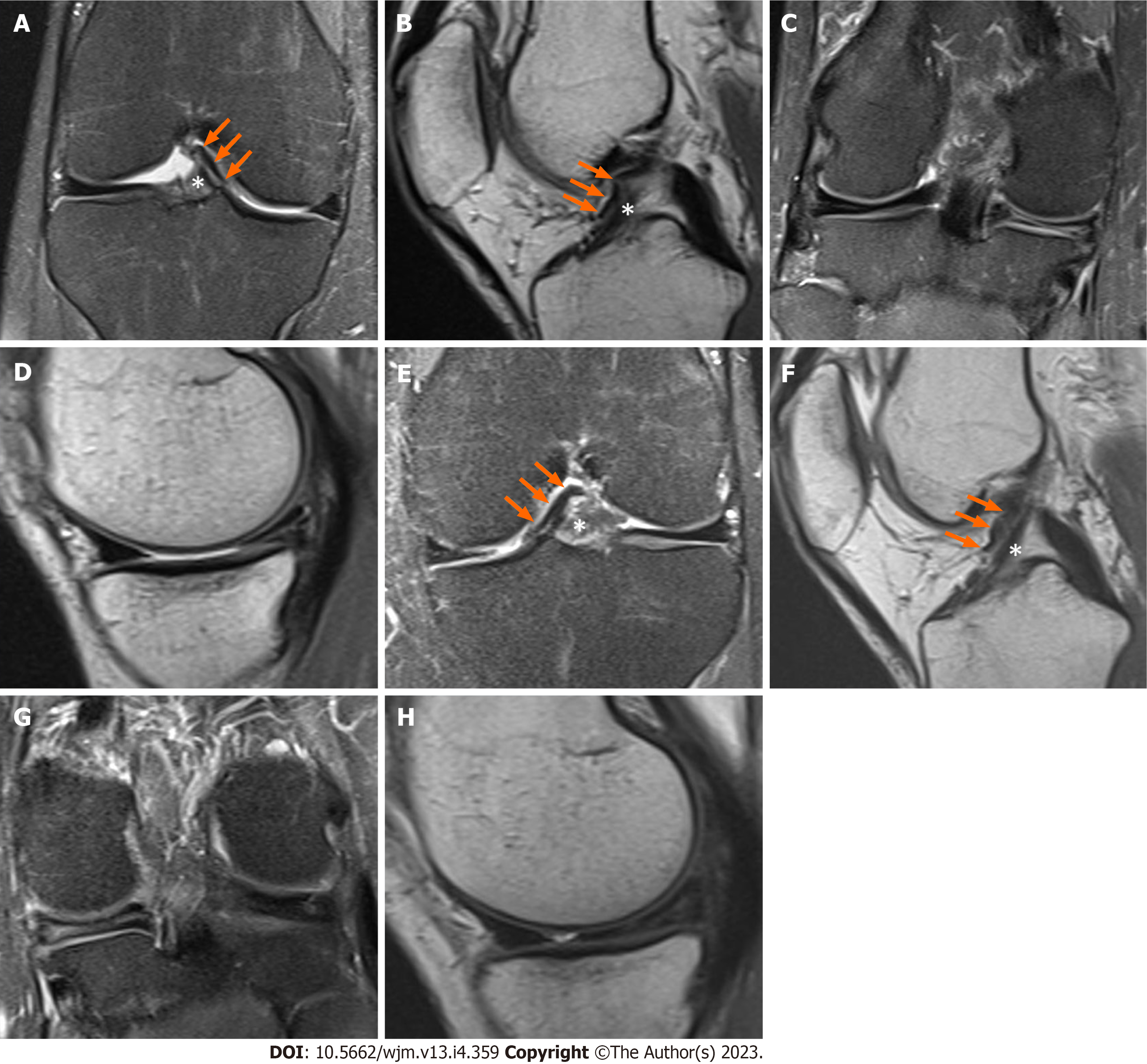
Figure 1 Magnetic resonance imaging images of the knee.
A-D: Magnetic resonance imaging (MRI) images of the right knee. Coronal T2-weighted fat-saturated image demonstrating the anteromedial meniscofemoral ligament (AMMFL) (green arrow) and the distal aspect of the anterior cruciate ligament (ACL) (white asterisk) (A); sagittal T1-weighted image showing the AMMFL (green arrow) running anteriorly to the ACL (white asterisk) (B); coronal T2-weighted fat-saturated and sagittal images showing the medial meniscus with a previous partial meniscectomy and tear of the posterior horn (C and D); E-H: MRI images of the left knee. Coronal T2-weighted fat-saturated image demonstrating the AMMFL (green arrow) and the distal aspect of the ACL (white asterisk) (E); sagittal T1-weighted image showing the AMMFL (green arrow) running anteriorly to the ACL (white asterisk) (F); coronal T2-weighted fat-saturated and sagittal images showing a tear of the posterior horn of the medial meniscus (G and H).
- Citation: Luco JB, Di Memmo D, Gomez Sicre V, Nicolino TI, Costa-Paz M, Astoul J, Garcia-Mansilla I. Clinical, imaging, arthroscopic, and histologic features of bilateral anteromedial meniscofemoral ligament: A case report. World J Methodol 2023; 13(4): 359-365
- URL: https://www.wjgnet.com/2222-0682/full/v13/i4/359.htm
- DOI: https://dx.doi.org/10.5662/wjm.v13.i4.359


