Copyright
©The Author(s) 2022.
World J Methodol. Mar 20, 2022; 12(2): 64-82
Published online Mar 20, 2022. doi: 10.5662/wjm.v12.i2.64
Published online Mar 20, 2022. doi: 10.5662/wjm.v12.i2.64
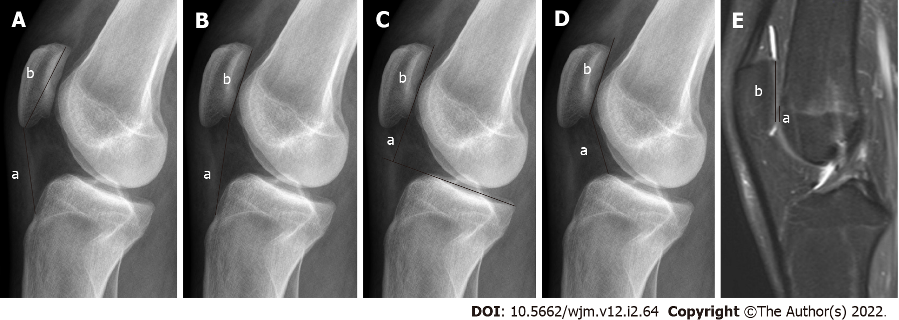
Figure 2 Radiography.
A: Insall-Salvati (IS) ratio (a: Patellar tendon length; b: Length of the patella); B: Modified IS ratio (a: The distance between the lower end of the articular face of the patella and TT; b: Length of the articular face of the patella); C: Blackburne-Peel ratio (a: The distance between the tibial plateau line and the lower pole of the patella joint surface; b: Length of the articular surface of the patella); D: Caton-Deschamps ratio (a: The distance between the inferior point of the patellar articular face and the anterior superior border of the tibia; b: Length of the articular surface of the patella); E: Magnetic resonance imaging, Patellotrochlear index (a: Length of the trochlear cartilage; b: Length of the patellar cartilage). These images are showing different measurement methods for evaluating the patellar height.
- Citation: Ormeci T, Turkten I, Sakul BU. Radiological evaluation of patellofemoral instability and possible causes of assessment errors. World J Methodol 2022; 12(2): 64-82
- URL: https://www.wjgnet.com/2222-0682/full/v12/i2/64.htm
- DOI: https://dx.doi.org/10.5662/wjm.v12.i2.64


