Copyright
©The Author(s) 2021.
World J Methodol. Jul 20, 2021; 11(4): 208-221
Published online Jul 20, 2021. doi: 10.5662/wjm.v11.i4.208
Published online Jul 20, 2021. doi: 10.5662/wjm.v11.i4.208
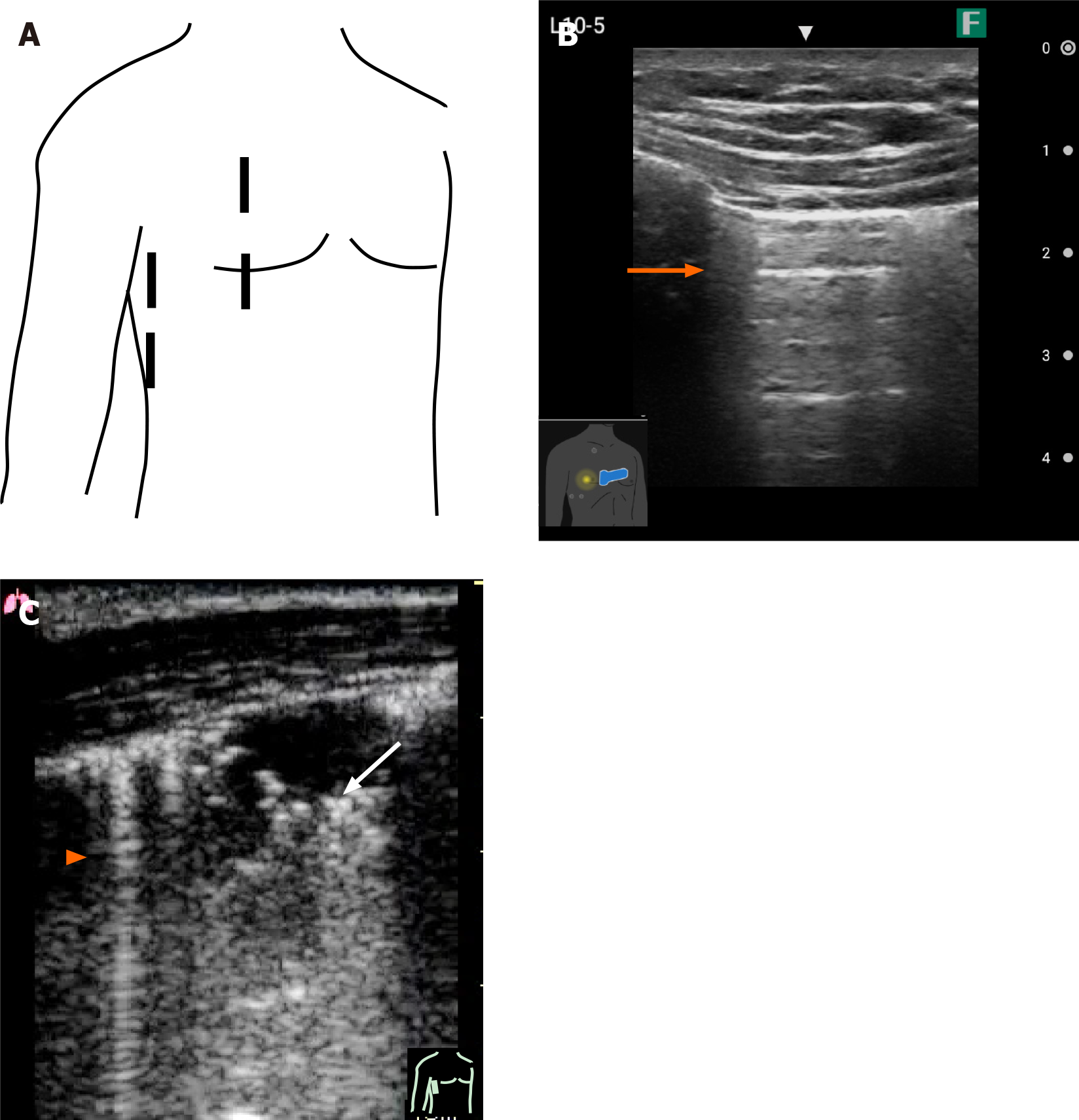
Figure 9 Pocket-sized ultrasound image of the pleura.
A: Basic scanning planes of the chest (right) by ultrasound; B: Ultrasound image of normal pleura showing a typical A line (orange arrow); C: Ultrasound image of B line (arrowhead) and consolidation of the lung (white arrow).
- Citation: Naganuma H, Ishida H. One-day seminar for residents for implementing abdominal pocket-sized ultrasound. World J Methodol 2021; 11(4): 208-221
- URL: https://www.wjgnet.com/2222-0682/full/v11/i4/208.htm
- DOI: https://dx.doi.org/10.5662/wjm.v11.i4.208


