Copyright
©The Author(s) 2021.
World J Methodol. Jul 20, 2021; 11(4): 208-221
Published online Jul 20, 2021. doi: 10.5662/wjm.v11.i4.208
Published online Jul 20, 2021. doi: 10.5662/wjm.v11.i4.208
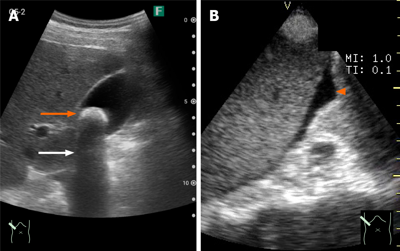
Figure 6 Pocket-sized ultrasound images of gallbladder stones and ascites.
A: A 1-cm stone; B: A small amount of ascites. A 1-cm stone (A) and a small amount of ascites (B) is clearly visualized as a strong echo (orange arrow) with acoustic shadowing (white arrow) in the former and an echo-free space in the latter (arrowhead).
- Citation: Naganuma H, Ishida H. One-day seminar for residents for implementing abdominal pocket-sized ultrasound. World J Methodol 2021; 11(4): 208-221
- URL: https://www.wjgnet.com/2222-0682/full/v11/i4/208.htm
- DOI: https://dx.doi.org/10.5662/wjm.v11.i4.208


