Copyright
©The Author(s) 2021.
World J Methodol. Jul 20, 2021; 11(4): 208-221
Published online Jul 20, 2021. doi: 10.5662/wjm.v11.i4.208
Published online Jul 20, 2021. doi: 10.5662/wjm.v11.i4.208
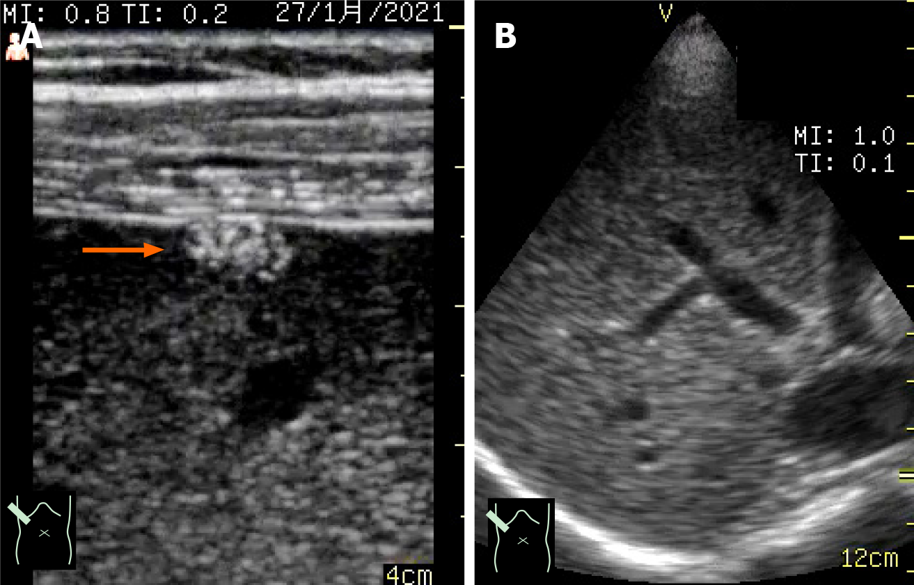
Figure 3 Ultrasound image by different probes on a pocket-sized ultrasound device.
A: High-frequency linear probe; B: 1.7-3.8 MHz sector probe. The linear probe is used to visualize superficial areas, and the sector probe is used for observing deep areas. In this case of a small liver tumor situated at the hepatic surface, the lesion was detected by the linear probe (→) but not the sector probe.
- Citation: Naganuma H, Ishida H. One-day seminar for residents for implementing abdominal pocket-sized ultrasound. World J Methodol 2021; 11(4): 208-221
- URL: https://www.wjgnet.com/2222-0682/full/v11/i4/208.htm
- DOI: https://dx.doi.org/10.5662/wjm.v11.i4.208


