Copyright
©The Author(s) 2021.
World J Methodol. Jul 20, 2021; 11(4): 208-221
Published online Jul 20, 2021. doi: 10.5662/wjm.v11.i4.208
Published online Jul 20, 2021. doi: 10.5662/wjm.v11.i4.208
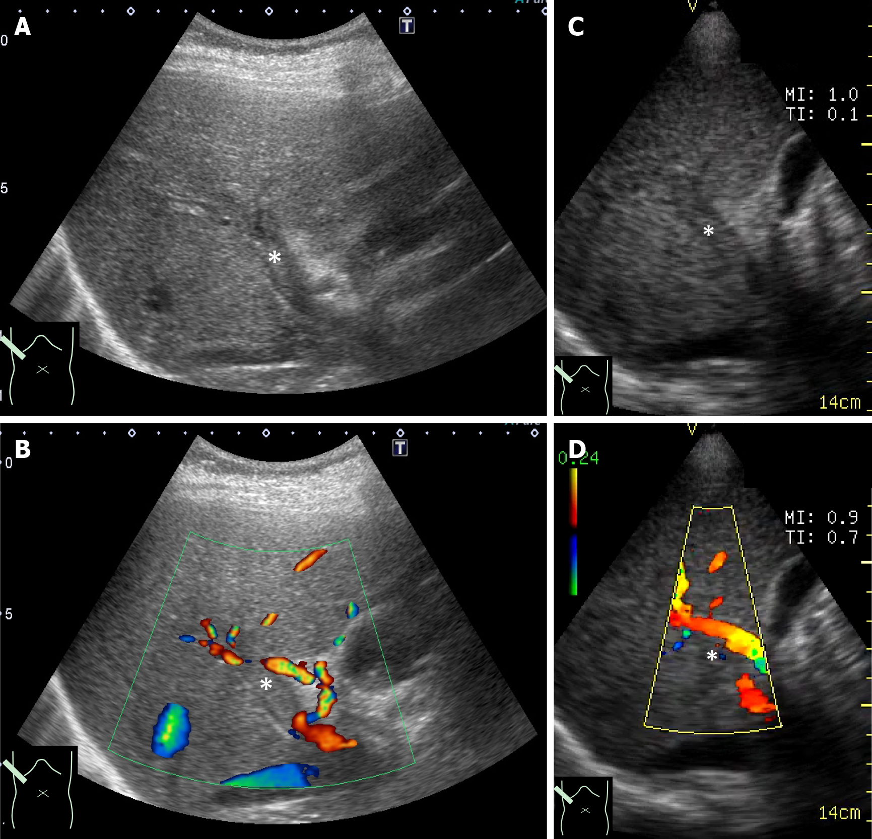
Figure 2 Simultaneous presentation of ultrasound images of a portal thrombus.
A: High-end ultrasound B mode; B: High-end ultrasound color Doppler; C: Pocket-sized ultrasound B mode; D: Pocket-sized ultrasound color Doppler. These comparisons are used to understand the difference in image quality between the two machines. It also confirms that pocket-sized ultrasound is sufficient for diagnoses. *: Thrombus in the portal vein.
- Citation: Naganuma H, Ishida H. One-day seminar for residents for implementing abdominal pocket-sized ultrasound. World J Methodol 2021; 11(4): 208-221
- URL: https://www.wjgnet.com/2222-0682/full/v11/i4/208.htm
- DOI: https://dx.doi.org/10.5662/wjm.v11.i4.208


