Copyright
©The Author(s) 2015.
World J Transl Med. Dec 12, 2015; 4(3): 113-122
Published online Dec 12, 2015. doi: 10.5528/wjtm.v4.i3.113
Published online Dec 12, 2015. doi: 10.5528/wjtm.v4.i3.113
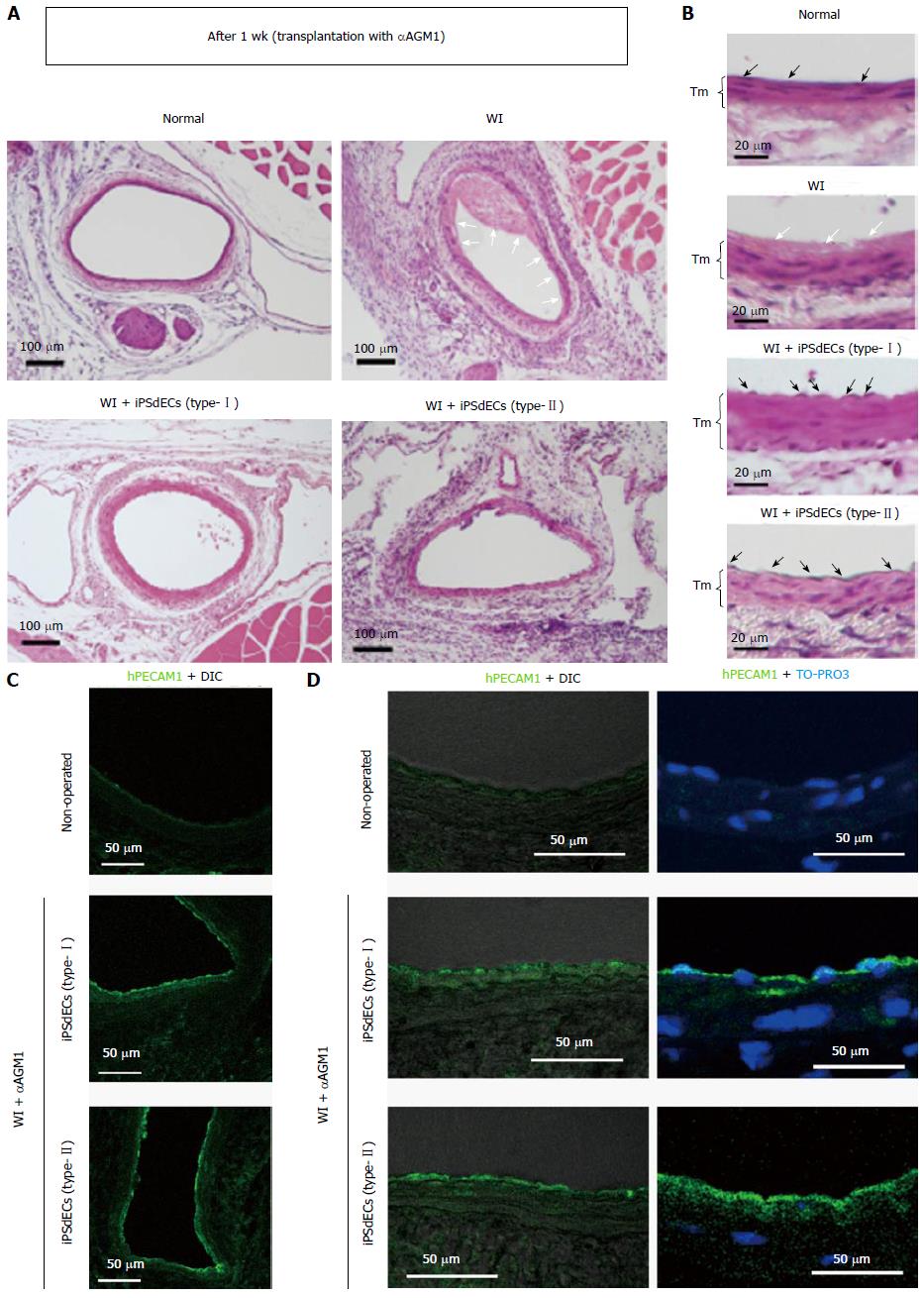
Figure 2 Histological analyses after one week.
A and B: WI-operated femoral arteries that were transplanted with type-I or type-II iPSdECs were examined after one week from PVVT in mice regularly administrated with asialo GM1 antibody αAGM1). Open arrows indicate fibrin deposits, closed arrows indicate nuclei of endothelial cells and Tm indicate tunica media; C and D: Con-focal microscopies of immunostained samples using anti-human PECAM1 antibody with differential interference contrast (DIC) (C) or nuclear counterstaining by TO-PRO3 (D). WI: Wire injury; iPSC: Induced pluripotent stem cell; VEC: Vascular endothelial cell; iPSdECs: iPSC-derived VECs; αAGM1: Anti-asialo GM1 monoclonal antibody.
- Citation: Nishio M, Nakahara M, Saeki K, Fujiu K, Iwata H, Manabe I, Yuo A, Saeki K. Pro- vs anti-stenotic capacities of type-I vs type-II human induced pluripotent-derived endothelial cells. World J Transl Med 2015; 4(3): 113-122
- URL: https://www.wjgnet.com/2220-6132/full/v4/i3/113.htm
- DOI: https://dx.doi.org/10.5528/wjtm.v4.i3.113


