Copyright
©The Author(s) 2015.
World J Transl Med. Dec 12, 2015; 4(3): 101-112
Published online Dec 12, 2015. doi: 10.5528/wjtm.v4.i3.101
Published online Dec 12, 2015. doi: 10.5528/wjtm.v4.i3.101
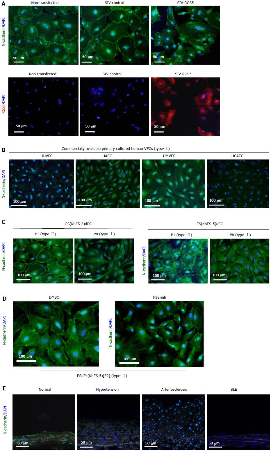
Figure 5 Effects of regulator of G-protein signaling 5 on the intracellular localization of N-cadherin.
A: Type-II VECs were infected with SIV vectors carrying either human RGS5 cDNA (SIV-RGS5) or inverted human RGS5 cDNA (SIV-control) and subjected to immunostaining using an anti-N-cadherin antibody (green) or anti-RGS5 antibody (red); B-E: Immunostaining studies using an anti-N-cadherin antibody (green) with nuclear counterstaining with DAPI (blue) were performed using commercially available primary cultured human VECs (type-I) (B), ESdECs at early (type-II) and late (type-I) passages (C), ESdECs at early passages (type-II) treated with 0.1% DMSO or a p38 inhibitor (10 μmol/L) (D) and clinical specimens (E). HCAEC: Human adult coronary arterial endothelial cells; HAEC: Human adult aortic endothelial cells; HMVEC: Human neonatal dermal microvascular endothelial cells; HUVEC: Human umbilical vein endothelial cells; ESdECs: Embryonic stem cells-derived vascular endothelial cells; RGS5: Regulator of G-protein signaling 5; DMSO: Dimethylsulfoxide; VECs: Vascular endothelial cells.
- Citation: Nakahara M, Nishio M, Saeki K, Yuo A, Saeki K. p38 mitogen-activated protein kinase regulates type-I vs type-II phenotyping of human vascular endothelial cells. World J Transl Med 2015; 4(3): 101-112
- URL: https://www.wjgnet.com/2220-6132/full/v4/i3/101.htm
- DOI: https://dx.doi.org/10.5528/wjtm.v4.i3.101


