Copyright
©The Author(s) 2018.
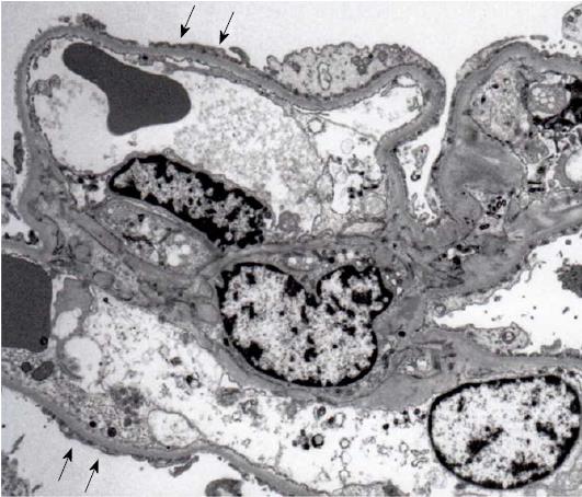
Figure 2 The results of electron microscopy.
Electron microscopy reveals a wide range of foot process effacement (arrows). No tubulo-reticular inclusions in the glomerular endothelium are seen and electron-dense deposits are absent (original magnification: 3000 ×).
- Citation: Goto D, Ohashi N, Takeda A, Fujigaki Y, Shimizu A, Yasuda H, Ohishi K. Case of human immunodeficiency virus infection presenting as a tip variant of focal segmental glomerulosclerosis: A case report and review of the literature. World J Nephrol 2018; 7(4): 90-95
- URL: https://www.wjgnet.com/2220-6124/full/v7/i4/90.htm
- DOI: https://dx.doi.org/10.5527/wjn.v7.i4.90


