Copyright
©The Author(s) 2017.
World J Nephrol. Nov 6, 2017; 6(6): 243-250
Published online Nov 6, 2017. doi: 10.5527/wjn.v6.i6.243
Published online Nov 6, 2017. doi: 10.5527/wjn.v6.i6.243
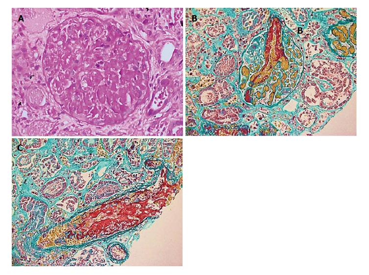
Figure 1 Finding of renal biopsy showed.
A: A glomerulus with extensive fibrin thrombi involvement, obliterating the capillary loops (Periodic Acid-Schiff stain, original magnification × 400); B: A glomerulus with an afferent arteriole with extensive red, fuchsinophilic material (fibrin) involvement (Masson trichrome stain, original magnification × 400); C: A small arteriole with luminal fibrin staining red (fuchsinophilic) (Masson trichrome stain, original magnification × 400).
- Citation: Almalki AH, Sadagah LF, Qureshi M, Maghrabi H, Algain A, Alsaeed A. Atypical hemolytic-uremic syndrome due to complement factor I mutation. World J Nephrol 2017; 6(6): 243-250
- URL: https://www.wjgnet.com/2220-6124/full/v6/i6/243.htm
- DOI: https://dx.doi.org/10.5527/wjn.v6.i6.243


