Copyright
©The Author(s) 2017.
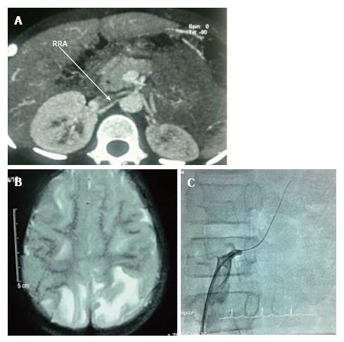
Figure 1 The angiography of three cases.
A: CT angiography shows narrowing of left renal artery with duplication of artery. Small left kidney with compensatory hypertrophy of right kidney; B: MRI brain shows posterior reversible encephalopathy syndrome; C: Digital subtraction angiography shows stenosed left renal artery. MRI: Magnetic resonance imaging; CT: Computed tomography.
- Citation: Mukherjee D, Sinha R, Akhtar MS, Saha AS. Hyponatremic hypertensive syndrome - a retrospective cohort study. World J Nephrol 2017; 6(1): 41-44
- URL: https://www.wjgnet.com/2220-6124/full/v6/i1/41.htm
- DOI: https://dx.doi.org/10.5527/wjn.v6.i1.41


