Copyright
©The Author(s) 2016.
World J Nephrol. Jul 6, 2016; 5(4): 339-357
Published online Jul 6, 2016. doi: 10.5527/wjn.v5.i4.339
Published online Jul 6, 2016. doi: 10.5527/wjn.v5.i4.339
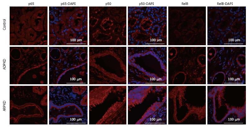
Figure 12 Immunofluorescence staining for p65, p50 and RelB (red) in human normal kidney cortex, and in autosomal dominant polycystic kidney disease and autosomal recessive polycystic kidney disease kidney cortex.
Also shown are corresponding DAPI-merged images with nuclei labeled using DAPI (blue). ADPKD: Autosomal dominant polycystic kidney disease; ARPKD: Autosomal recessive polycystic kidney disease; LPK: Lewis polycystic kidney.
- Citation: Ta MHT, Schwensen KG, Liuwantara D, Huso DL, Watnick T, Rangan GK. Constitutive renal Rel/nuclear factor-κB expression in Lewis polycystic kidney disease rats. World J Nephrol 2016; 5(4): 339-357
- URL: https://www.wjgnet.com/2220-6124/full/v5/i4/339.htm
- DOI: https://dx.doi.org/10.5527/wjn.v5.i4.339


