Copyright
©The Author(s) 2016.
World J Nephrol. Jul 6, 2016; 5(4): 339-357
Published online Jul 6, 2016. doi: 10.5527/wjn.v5.i4.339
Published online Jul 6, 2016. doi: 10.5527/wjn.v5.i4.339
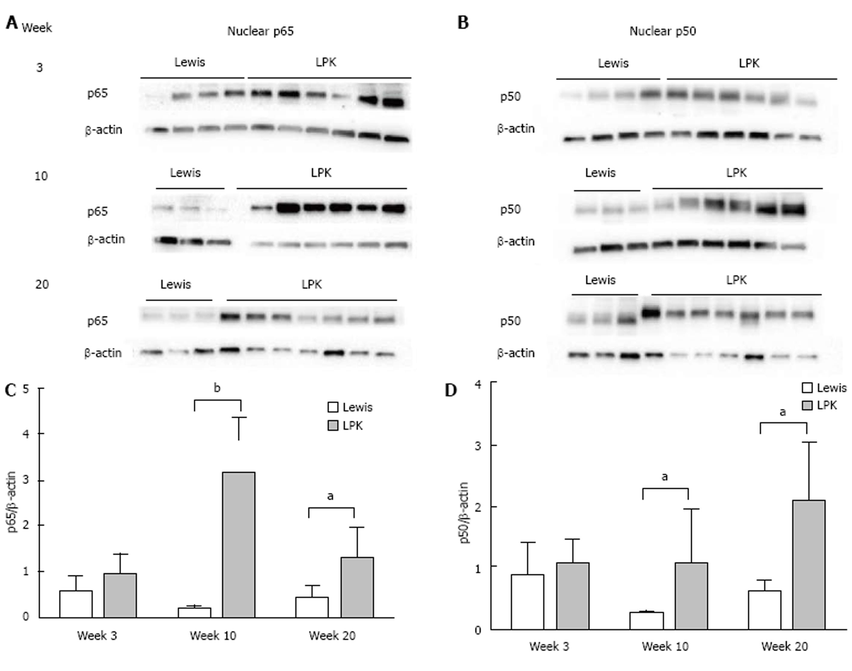
Figure 8 Western blotting for nuclear factor-κB p65 and p50 proteins in Lewis and Lewis polycystic kidney.
Immunoblotting was performed for (A) nuclear p65, and (B) nuclear p50, in Lewis and LPK kidney tissue from weeks 3, 10 and 20. Densitometry of immunoblots was quantified for (C) p65 and (D) p50. aP < 0.05 vs Lewis for the corresponding timepoint; bP < 0.01 vs Lewis for the corresponding timepoint. LPK: Lewis polycystic kidney.
- Citation: Ta MHT, Schwensen KG, Liuwantara D, Huso DL, Watnick T, Rangan GK. Constitutive renal Rel/nuclear factor-κB expression in Lewis polycystic kidney disease rats. World J Nephrol 2016; 5(4): 339-357
- URL: https://www.wjgnet.com/2220-6124/full/v5/i4/339.htm
- DOI: https://dx.doi.org/10.5527/wjn.v5.i4.339


