Copyright
©The Author(s) 2015.
World J Nephrol. May 6, 2015; 4(2): 185-195
Published online May 6, 2015. doi: 10.5527/wjn.v4.i2.185
Published online May 6, 2015. doi: 10.5527/wjn.v4.i2.185
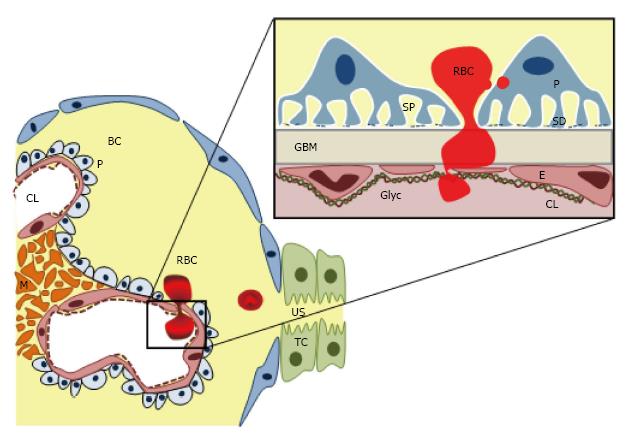
Figure 2 Glomerular filtration barrier structure and red blood cell egression leading to haematuria.
CL: Capillary lumen; BC: Bowman’s capsule; E: Endothelial cell; GBM: Glomerular basement membrane; Gly: Glycosaminoglicans; M: Mesangium; P: Podocyte; RBC: Red blood cell; SD: Slit diaphragm; SP: Subpodocyte space; TC: Tubular cell; US: Urinary space.
- Citation: Yuste C, Gutierrez E, Sevillano AM, Rubio-Navarro A, Amaro-Villalobos JM, Ortiz A, Egido J, Praga M, Moreno JA. Pathogenesis of glomerular haematuria. World J Nephrol 2015; 4(2): 185-195
- URL: https://www.wjgnet.com/2220-6124/full/v4/i2/185.htm
- DOI: https://dx.doi.org/10.5527/wjn.v4.i2.185


