Copyright
©The Author(s) 2015.
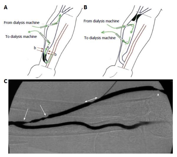
Figure 1 Schematic of the locations of (A) inflow stenosis, and (B) outflow lesion with respect to the anastomosis and cannulation sites[27].
The (green) arrows in A and B represent the direction of blood flow within the arteriovenous fistulas (AVF) and the dialysis needles; B: Depicts a recirculation condition under which due to significant outflow stenosis the blood flow from dialysis machine returns back to dialyzer; C: An angiographic picture of an AVF with multiple inflow stenoses (single head arrows) and outflow stenosis (double head arrow and the arrow head). Reprinted from Asif et al[25] and Fahrtash et al[27], with permission.
- Citation: Rajabi-Jaghargh E, Banerjee RK. Combined functional and anatomical diagnostic endpoints for assessing arteriovenous fistula dysfunction. World J Nephrol 2015; 4(1): 6-18
- URL: https://www.wjgnet.com/2220-6124/full/v4/i1/6.htm
- DOI: https://dx.doi.org/10.5527/wjn.v4.i1.6


