Copyright
©2014 Baishideng Publishing Group Inc.
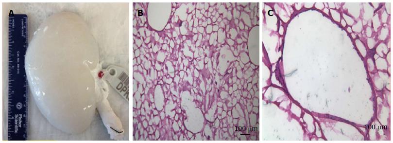
Figure 3 A decellularized pig kidney scaffold and its extra cellular matrix after decellularization.
A: Decellularized pig kidney scaffold; B: Hematoxylin and eosin staining of the decellularized pig kidney scaffold shows a decellularized extracellular matrix (× 200); C: Hematoxylin and eosin staining of the decellularized pig kidney scaffold shows a decellularized extracellular matrix (× 400). Permission of Wake Forest Institute for Regenerative Medicine.
- Citation: Zambon JP, Magalhaes RS, Ko I, Ross CL, Orlando G, Peloso A, Atala A, Yoo JJ. Kidney regeneration: Where we are and future perspectives. World J Nephrol 2014; 3(3): 24-30
- URL: https://www.wjgnet.com/2220-6124/full/v3/i3/24.htm
- DOI: https://dx.doi.org/10.5527/wjn.v3.i3.24


