Copyright
©The Author(s) 2025.
World J Nephrol. Jun 25, 2025; 14(2): 105374
Published online Jun 25, 2025. doi: 10.5527/wjn.v14.i2.105374
Published online Jun 25, 2025. doi: 10.5527/wjn.v14.i2.105374
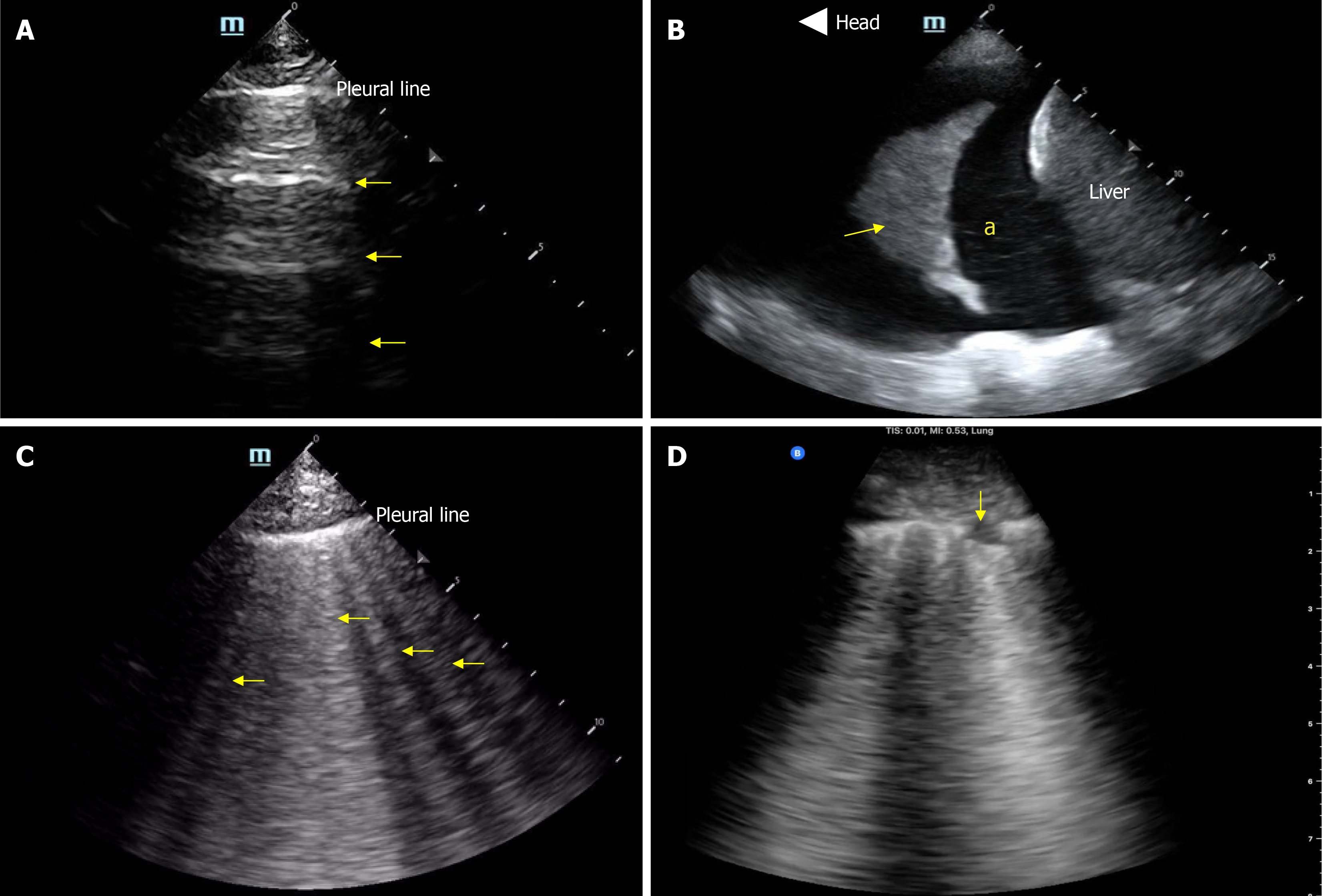
Figure 2 Basic lung ultrasound findings.
A: Normal lung ultrasound demonstrating A-lines (horizontal hyperechoic artifacts); B: Pleural effusion (“a”) appearing as an anechoic area above the liver. Arrow points to atelectatic lung; C: B-lines-vertical hyperechoic artifacts emerging from the pleural line indicative of interstitial thickening (typically from fluid); D: Interstitial pneumonia with confluent B-lines and an irregular pleural line. Arrow points to subpleural consolidation.
- Citation: Diniz H, Ferreira F, Koratala A. Point-of-care ultrasonography in nephrology: Growing applications, misconceptions and future outlook. World J Nephrol 2025; 14(2): 105374
- URL: https://www.wjgnet.com/2220-6124/full/v14/i2/105374.htm
- DOI: https://dx.doi.org/10.5527/wjn.v14.i2.105374


