Copyright
©The Author(s) 2025.
World J Nephrol. Jun 25, 2025; 14(2): 103027
Published online Jun 25, 2025. doi: 10.5527/wjn.v14.i2.103027
Published online Jun 25, 2025. doi: 10.5527/wjn.v14.i2.103027
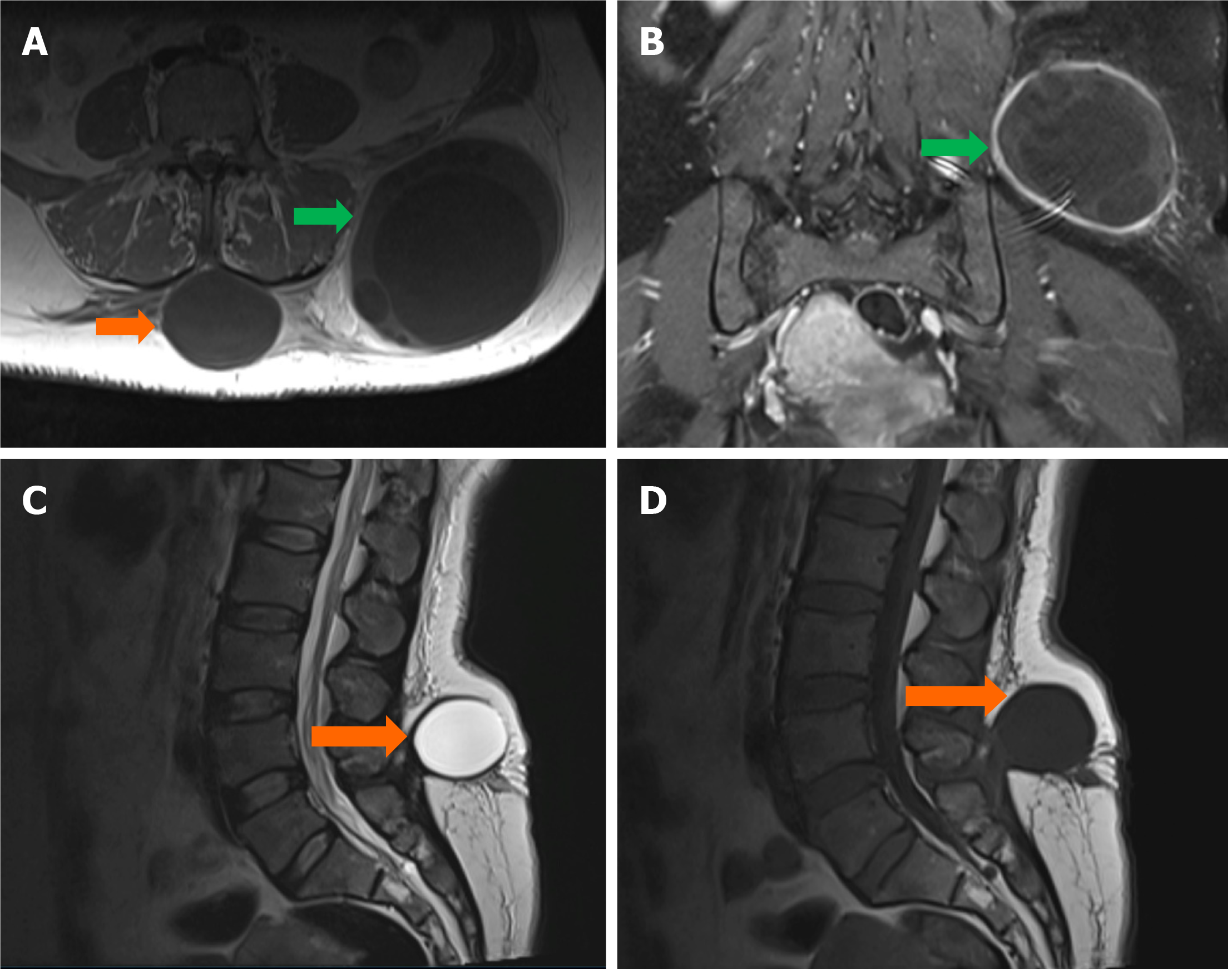
Figure 3 Lumbar region magnetic resonance imaging of multiloculated cystic lesions in the subcutaneous fat tissue.
A: Axial T1W image reveals two distinct multiloculated cystic lesions in the lumbar subcutaneous fat tissue, one located in the midline (orange arrows) and the other on the left lateral side (green arrows); B: Coronal post-contrast fat-saturated T1W image shows contrast enhancement in both the septal and mural components of the lesions; C: Sagittal T2W image demonstrates the cystic nature of the lesions; D: Sagittal T1W image further characterizes the lesions with visible enhancement in their septal and mural components.
- Citation: Celik AS, Yosunkaya H, Yayilkan Ozyilmaz A, Memis KB, Aydin S. Echinococcus granulosus in atypical localizations: Five case reports. World J Nephrol 2025; 14(2): 103027
- URL: https://www.wjgnet.com/2220-6124/full/v14/i2/103027.htm
- DOI: https://dx.doi.org/10.5527/wjn.v14.i2.103027


